 Movie
Movie Controller

[English] 日本語
 Yorodumi
- PDB-7tzt: Crystal structure of the E. coli thiM riboswitch in complex with ... -
+ Open data

- Basic information
| Entry | Database: PDB / ID: 7tzt | ||||||
|---|---|---|---|---|---|---|---|
| Title | Crystal structure of the E. coli thiM riboswitch in complex with N1,N1-dimethyl-N2-(quinoxalin-6-ylmethyl)ethane-1,2-diamine (linked compound 37) | ||||||
 Components Components | RNA (79-MER) | ||||||
 Keywords Keywords | RNA / thiM TPP riboswitch | ||||||
| Function / homology | 6-methylquinoxaline / : / RNA / RNA (> 10) Function and homology information Function and homology information | ||||||
| Biological species |  | ||||||
| Method |  X-RAY DIFFRACTION / SYNCHROTRON / MOLECULAR REPLACEMENT / Resolution: 2.96 Å X-RAY DIFFRACTION / SYNCHROTRON / MOLECULAR REPLACEMENT / Resolution: 2.96 Å | ||||||
 Authors Authors | Nuthanakanti, A. / Serganov, A. | ||||||
| Funding support |  United States, 1items United States, 1items
| ||||||
 Citation Citation | Journal: Proc.Natl.Acad.Sci.USA / Year: 2022 Title: SHAPE-enabled fragment-based ligand discovery for RNA. Authors: Zeller, M.J. / Favorov, O. / Li, K. / Nuthanakanti, A. / Hussein, D. / Michaud, A. / Lafontaine, D.A. / Busan, S. / Serganov, A. / Aube, J. / Weeks, K.M. | ||||||
| History |
|
- Structure visualization
| Structure viewer | Molecule: MolmilJmol/JSmol |
|---|
- Downloads & links
-Download
| PDBx/mmCIF format | 7tzt.cif.gz | 102.4 KB | Display | PDBx/mmCIF format |
|---|---|---|---|---|
| PDB format | pdb7tzt.ent.gz | 74.2 KB | Display | PDB format |
| PDBx/mmJSON format | 7tzt.json.gz | Tree view | PDBx/mmJSON format | |
| Others |  Other downloads Other downloads |
-Validation report
| Arichive directory | https://data.pdbj.org/pub/pdb/validation_reports/tz/7tztftp://data.pdbj.org/pub/pdb/validation_reports/tz/7tzt | HTTPS FTP |
|---|
-Related structure data
| Related structure data |  7tzrC 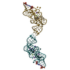 7tzsC 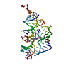 7tzuC  2hojS S: Starting model for refinement C: citing same article ( |
|---|---|
| Similar structure data |
-Links
 PDBj
PDBj


- Assembly
| Deposited unit | 
| ||||||||||||
|---|---|---|---|---|---|---|---|---|---|---|---|---|---|
| 1 |
| ||||||||||||
| Unit cell |
|
-Components
| #1: RNA chain | Mass: 26798.936 Da / Num. of mol.: 1 / Source method: obtained synthetically / Source: (synth.) | ||||||
|---|---|---|---|---|---|---|---|
| #2: Chemical |   Mass: 54.938 Da / Num. of mol.: 2 / Source method: obtained synthetically / Formula: Mn Mass: 54.938 Da / Num. of mol.: 2 / Source method: obtained synthetically / Formula: Mn#3: Chemical | ChemComp-MG / |   Mass: 24.305 Da / Num. of mol.: 1 / Source method: obtained synthetically / Formula: Mg Mass: 24.305 Da / Num. of mol.: 1 / Source method: obtained synthetically / Formula: Mg#4: Chemical | ChemComp-KXC / |   Mass: 230.309 Da / Num. of mol.: 1 / Source method: obtained synthetically / Formula: C13H18N4 / Feature type: SUBJECT OF INVESTIGATION Mass: 230.309 Da / Num. of mol.: 1 / Source method: obtained synthetically / Formula: C13H18N4 / Feature type: SUBJECT OF INVESTIGATIONHas ligand of interest | Y | |
-Experimental details
-Experiment
| Experiment | Method: X-RAY DIFFRACTION / Number of used crystals: 1 |
|---|
- Sample preparation
| Crystal | Density Matthews: 2.06 Å3/Da / Density % sol: 40.36 % / Description: large trigonal shaped crystals |
|---|---|
| Crystal grow | Temperature: 291 K / Method: vapor diffusion, hanging drop Details: The RNA (0.15 mM) was incubated in a buffer containing 5 mM Tris-HCl, pH 8.0, 3 mM MgCl2, 10 mM NaCl, 0.1 M KCl, and 0.5 mM spermine with 1.0 mM compound 38. Reservoir solution was 50 mM Bis- ...Details: The RNA (0.15 mM) was incubated in a buffer containing 5 mM Tris-HCl, pH 8.0, 3 mM MgCl2, 10 mM NaCl, 0.1 M KCl, and 0.5 mM spermine with 1.0 mM compound 38. Reservoir solution was 50 mM Bis-Tris, pH 6.5, 0.5 M ammonium chloride, 15 mm MnCl2, and 28% (v/v) PEG2000 PH range: 6.5-8.0 |
-Data collection
| Diffraction | Mean temperature: 100 K / Serial crystal experiment: N |
|---|---|
| Diffraction source | Source: SYNCHROTRON / Site: APS / Beamline: 24-ID-C / Wavelength: 0.9791 Å |
| Detector | Type: DECTRIS EIGER2 X 16M / Detector: PIXEL / Date: Mar 21, 2021 |
| Radiation | Protocol: SINGLE WAVELENGTH / Monochromatic (M) / Laue (L): M / Scattering type: x-ray |
| Radiation wavelength | Wavelength: 0.9791 Å / Relative weight: 1 |
| Reflection | Resolution: 2.93→50 Å / Num. obs: 4715 / % possible obs: 99.9 % / Redundancy: 10 % / Biso Wilson estimate: 97.5 Å2 / CC1/2: 0.995 / Rmerge(I) obs: 0.13 / Rpim(I) all: 0.044 / Net I/σ(I): 304.8 |
| Reflection shell | Resolution: 2.93→2.98 Å / Redundancy: 10.6 % / Rmerge(I) obs: 3.612 / Mean I/σ(I) obs: 5.5 / Num. unique obs: 238 / CC1/2: 0.637 / Rpim(I) all: 1.14 / % possible all: 100 |
- Processing
| Software |
| |||||||||||||||||||||||||||||||||||||||||||||||||
|---|---|---|---|---|---|---|---|---|---|---|---|---|---|---|---|---|---|---|---|---|---|---|---|---|---|---|---|---|---|---|---|---|---|---|---|---|---|---|---|---|---|---|---|---|---|---|---|---|---|---|
| Refinement | Method to determine structure: MOLECULAR REPLACEMENT Starting model: 2HOJ Resolution: 2.96→47 Å / SU ML: 0.3197 / Cross valid method: FREE R-VALUE / σ(F): 1.36 / Phase error: 36.9635 Stereochemistry target values: GeoStd + Monomer Library + CDL v1.2
| |||||||||||||||||||||||||||||||||||||||||||||||||
| Solvent computation | Shrinkage radii: 0.9 Å / VDW probe radii: 1.11 Å / Solvent model: FLAT BULK SOLVENT MODEL | |||||||||||||||||||||||||||||||||||||||||||||||||
| Displacement parameters | Biso mean: 115.66 Å2 | |||||||||||||||||||||||||||||||||||||||||||||||||
| Refinement step | Cycle: LAST / Resolution: 2.96→47 Å
| |||||||||||||||||||||||||||||||||||||||||||||||||
| Refine LS restraints |
| |||||||||||||||||||||||||||||||||||||||||||||||||
| LS refinement shell |
| |||||||||||||||||||||||||||||||||||||||||||||||||
| Refinement TLS params. | Method: refined / Origin x: -8.30212374089 Å / Origin y: 16.841211741 Å / Origin z: 5.43754207531 Å
| |||||||||||||||||||||||||||||||||||||||||||||||||
| Refinement TLS group | Selection details: all |
