Movie
Movie Controller

[English] 日本語
 Yorodumi
- PDB-7ty7: Cryo-EM structure of human Anion Exchanger 1 bound to Bicarbonate -
+ Open data

- Basic information
| Entry | Database: PDB / ID: 7ty7 | ||||||||||||
|---|---|---|---|---|---|---|---|---|---|---|---|---|---|
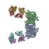
| Title | Cryo-EM structure of human Anion Exchanger 1 bound to Bicarbonate | ||||||||||||
 Components Components | Band 3 anion transport protein | ||||||||||||
 Keywords Keywords | TRANSPORT PROTEIN / Transmembrane | ||||||||||||
| Function / homology |  Function and homology information Function and homology informationresponse to increased oxygen levels / pH elevation / Defective SLC4A1 causes hereditary spherocytosis type 4 (HSP4), distal renal tubular acidosis (dRTA) and dRTA with hemolytic anemia (dRTA-HA) / negative regulation of urine volume / Bicarbonate transporters / ankyrin-1 complex / intracellular monoatomic ion homeostasis / monoatomic anion transmembrane transporter activity / plasma membrane phospholipid scrambling / chloride:bicarbonate antiporter activity ...response to increased oxygen levels / pH elevation / Defective SLC4A1 causes hereditary spherocytosis type 4 (HSP4), distal renal tubular acidosis (dRTA) and dRTA with hemolytic anemia (dRTA-HA) / negative regulation of urine volume / Bicarbonate transporters / ankyrin-1 complex / intracellular monoatomic ion homeostasis / monoatomic anion transmembrane transporter activity / plasma membrane phospholipid scrambling / chloride:bicarbonate antiporter activity / solute:inorganic anion antiporter activity / bicarbonate transmembrane transporter activity / bicarbonate transport / monoatomic anion transport / chloride transmembrane transporter activity / chloride transport / ankyrin binding / hemoglobin binding / negative regulation of glycolytic process through fructose-6-phosphate / cortical cytoskeleton / erythrocyte development / protein-membrane adaptor activity / chloride transmembrane transport / regulation of intracellular pH / protein localization to plasma membrane / Erythrocytes take up oxygen and release carbon dioxide / Erythrocytes take up carbon dioxide and release oxygen / transmembrane transport / Z disc / cytoplasmic side of plasma membrane / blood coagulation / blood microparticle / basolateral plasma membrane / protein homodimerization activity / extracellular exosome / membrane / plasma membrane Similarity search - Function | ||||||||||||
| Biological species |  Homo sapiens (human) Homo sapiens (human) | ||||||||||||
| Method | ELECTRON MICROSCOPY / single particle reconstruction / cryo EM / Resolution: 3.37 Å | ||||||||||||
 Authors Authors | Capper, M.J. / Mathiharan, Y.K. / Yang, S. / Stone, A.C. / Wacker, D. | ||||||||||||
| Funding support |  United States, 3items United States, 3items
| ||||||||||||
 Citation Citation | Journal: Nat Struct Mol Biol / Year: 2023 Title: Substrate binding and inhibition of the anion exchanger 1 transporter. Authors: Michael J Capper / Shifan Yang / Alexander C Stone / Sezen Vatansever / Gregory Zilberg / Yamuna Kalyani Mathiharan / Raul Habib / Keino Hutchinson / Yihan Zhao / Avner Schlessinger / Mihaly ...Authors: Michael J Capper / Shifan Yang / Alexander C Stone / Sezen Vatansever / Gregory Zilberg / Yamuna Kalyani Mathiharan / Raul Habib / Keino Hutchinson / Yihan Zhao / Avner Schlessinger / Mihaly Mezei / Roman Osman / Bin Zhang / Daniel Wacker / Abstract: Anion exchanger 1 (AE1), a member of the solute carrier (SLC) family, is the primary bicarbonate transporter in erythrocytes, regulating pH levels and CO transport between lungs and tissues. Previous ...Anion exchanger 1 (AE1), a member of the solute carrier (SLC) family, is the primary bicarbonate transporter in erythrocytes, regulating pH levels and CO transport between lungs and tissues. Previous studies characterized its role in erythrocyte structure and provided insight into transport regulation. However, key questions remain regarding substrate binding and transport, mechanisms of drug inhibition and modulation by membrane components. Here we present seven cryo-EM structures in apo, bicarbonate-bound and inhibitor-bound states. These, combined with uptake and computational studies, reveal important molecular features of substrate recognition and transport, and illuminate sterol binding sites, to elucidate distinct inhibitory mechanisms of research chemicals and prescription drugs. We further probe the substrate binding site via structure-based ligand screening, identifying an AE1 inhibitor. Together, our findings provide insight into mechanisms of solute carrier transport and inhibition. | ||||||||||||
| History |
|
- Structure visualization
| Structure viewer | Molecule: MolmilJmol/JSmol |
|---|
- Downloads & links
-Download
| PDBx/mmCIF format | 7ty7.cif.gz | 239.8 KB | Display | PDBx/mmCIF format |
|---|---|---|---|---|
| PDB format | pdb7ty7.ent.gz | 179.6 KB | Display | PDB format |
| PDBx/mmJSON format | 7ty7.json.gz | Tree view | PDBx/mmJSON format | |
| Others |  Other downloads Other downloads |
-Validation report
| Arichive directory | https://data.pdbj.org/pub/pdb/validation_reports/ty/7ty7ftp://data.pdbj.org/pub/pdb/validation_reports/ty/7ty7 | HTTPS FTP |
|---|
-Related structure data
| Related structure data |  26168MC 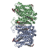 7ty4C 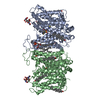 7ty6C 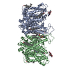 7ty8C 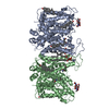 7tyaC 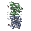 8t6uC 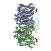 8t6vC M: map data used to model this data C: citing same article ( |
|---|---|
| Similar structure data |


-Links
 PDBj
PDBj


- Assembly
| Deposited unit | 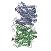
| ||||||||||||||||||||||||||||||
|---|---|---|---|---|---|---|---|---|---|---|---|---|---|---|---|---|---|---|---|---|---|---|---|---|---|---|---|---|---|---|---|
| 1 |
| ||||||||||||||||||||||||||||||
| Noncrystallographic symmetry (NCS) | NCS domain:
NCS domain segments: Component-ID: 1 / Ens-ID: 1 / Beg auth comp-ID: ASP / Beg label comp-ID: ASP / End auth comp-ID: ALA / End label comp-ID: ALA / Auth seq-ID: 369 - 891 / Label seq-ID: 369 - 891
NCS ensembles : (Details: Global NCS restraints between domains: 1 2) NCS oper:
|
-Components
-Protein / Sugars , 2 types, 4 molecules AB
| #1: Protein | Mass: 101883.859 Da / Num. of mol.: 2 Source method: isolated from a genetically manipulated source Source: (gene. exp.) Homo sapiens (human) / Gene: SLC4A1, AE1, DI, EPB3 / Plasmid: pFB / Cell line (production host): Sf9 / Production host:   Spodoptera frugiperda (fall armyworm) / References: UniProt: P02730 Spodoptera frugiperda (fall armyworm) / References: UniProt: P02730#2: Polysaccharide | Source method: isolated from a genetically manipulated source |
|---|
-Non-polymers , 4 types, 31 molecules 


| #3: Chemical |  Mass: 61.017 Da / Num. of mol.: 2 / Source method: obtained synthetically / Formula: CHO3 / Feature type: SUBJECT OF INVESTIGATION / Comment: pH buffer*YM Mass: 61.017 Da / Num. of mol.: 2 / Source method: obtained synthetically / Formula: CHO3 / Feature type: SUBJECT OF INVESTIGATION / Comment: pH buffer*YM#4: Chemical | ChemComp-CLR /  Mass: 386.654 Da / Num. of mol.: 7 / Source method: obtained synthetically / Formula: C27H46O Mass: 386.654 Da / Num. of mol.: 7 / Source method: obtained synthetically / Formula: C27H46O#5: Chemical |  Mass: 790.145 Da / Num. of mol.: 2 / Source method: obtained synthetically / Formula: C44H88NO8P / Comment: phospholipid*YM Mass: 790.145 Da / Num. of mol.: 2 / Source method: obtained synthetically / Formula: C44H88NO8P / Comment: phospholipid*YM#6: Water | ChemComp-HOH / | Mass: 18.015 Da / Num. of mol.: 20 / Source method: isolated from a natural source / Formula: H2O |
|---|
-Details
| Has ligand of interest | Y |
|---|---|
| Has protein modification | Y |
-Experimental details
-Experiment
| Experiment | Method: ELECTRON MICROSCOPY |
|---|---|
| EM experiment | Aggregation state: PARTICLE / 3D reconstruction method: single particle reconstruction |
- Sample preparation
| Component | Name: Dimeric anion exchanger 1 (SLC4A1) / Type: COMPLEX / Entity ID: #1 / Source: RECOMBINANT | ||||||||||||||||||||||||
|---|---|---|---|---|---|---|---|---|---|---|---|---|---|---|---|---|---|---|---|---|---|---|---|---|---|
| Molecular weight | Value: 0.203 MDa / Experimental value: NO | ||||||||||||||||||||||||
| Source (natural) | Organism: Homo sapiens (human) | ||||||||||||||||||||||||
| Source (recombinant) | Organism: Spodoptera frugiperda (fall armyworm) / Strain: Sf9 / Plasmid: pFB | ||||||||||||||||||||||||
| Buffer solution | pH: 7.5 | ||||||||||||||||||||||||
| Buffer component |
| ||||||||||||||||||||||||
| Specimen | Conc.: 5 mg/ml / Embedding applied: NO / Shadowing applied: NO / Staining applied: NO / Vitrification applied: YES Details: The sample was monodisperse following gel filtration and immediately concentrated for grid preparation | ||||||||||||||||||||||||
| Specimen support | Grid material: COPPER / Grid mesh size: 300 divisions/in. / Grid type: Quantifoil R1.2/1.3 | ||||||||||||||||||||||||
| Vitrification | Instrument: LEICA EM GP / Cryogen name: ETHANE / Humidity: 95 % / Chamber temperature: 285 K Details: Blot force 3 for 3-5 seconds was used and subsequent grids were screened for ice thickness prior to data collection |
- Electron microscopy imaging
| Experimental equipment |  Model: Titan Krios / Image courtesy: FEI Company |
|---|---|
| Microscopy | Model: TFS KRIOS |
| Electron gun | Electron source:  FIELD EMISSION GUN / Accelerating voltage: 300 kV / Illumination mode: FLOOD BEAM FIELD EMISSION GUN / Accelerating voltage: 300 kV / Illumination mode: FLOOD BEAM |
| Electron lens | Mode: BRIGHT FIELD / Nominal magnification: 64000 X / Nominal defocus max: 1800 nm / Nominal defocus min: 500 nm / Cs: 2.7 mm |
| Specimen holder | Cryogen: NITROGEN / Specimen holder model: FEI TITAN KRIOS AUTOGRID HOLDER |
| Image recording | Electron dose: 59.99 e/Å2 / Film or detector model: GATAN K3 (6k x 4k) / Num. of grids imaged: 1 / Num. of real images: 5256 |
- Processing
| Software | Name: REFMAC / Version: 5.8.0267 / Classification: refinement | ||||||||||||||||||||||||||||||||||||||||||||||||||||||||||||||||||||||||||||||||||||||||||||||||||||||||||
|---|---|---|---|---|---|---|---|---|---|---|---|---|---|---|---|---|---|---|---|---|---|---|---|---|---|---|---|---|---|---|---|---|---|---|---|---|---|---|---|---|---|---|---|---|---|---|---|---|---|---|---|---|---|---|---|---|---|---|---|---|---|---|---|---|---|---|---|---|---|---|---|---|---|---|---|---|---|---|---|---|---|---|---|---|---|---|---|---|---|---|---|---|---|---|---|---|---|---|---|---|---|---|---|---|---|---|---|
| EM software |
| ||||||||||||||||||||||||||||||||||||||||||||||||||||||||||||||||||||||||||||||||||||||||||||||||||||||||||
| CTF correction | Type: PHASE FLIPPING AND AMPLITUDE CORRECTION | ||||||||||||||||||||||||||||||||||||||||||||||||||||||||||||||||||||||||||||||||||||||||||||||||||||||||||
| Particle selection | Num. of particles selected: 2660401 | ||||||||||||||||||||||||||||||||||||||||||||||||||||||||||||||||||||||||||||||||||||||||||||||||||||||||||
| Symmetry | Point symmetry: C2 (2 fold cyclic) | ||||||||||||||||||||||||||||||||||||||||||||||||||||||||||||||||||||||||||||||||||||||||||||||||||||||||||
| 3D reconstruction | Resolution: 3.37 Å / Resolution method: FSC 0.143 CUT-OFF / Num. of particles: 173471 / Num. of class averages: 1 / Symmetry type: POINT | ||||||||||||||||||||||||||||||||||||||||||||||||||||||||||||||||||||||||||||||||||||||||||||||||||||||||||
| Atomic model building | Space: RECIPROCAL | ||||||||||||||||||||||||||||||||||||||||||||||||||||||||||||||||||||||||||||||||||||||||||||||||||||||||||
| Atomic model building | PDB-ID: 4YZF Accession code: 4YZF / Source name: PDB / Type: experimental model | ||||||||||||||||||||||||||||||||||||||||||||||||||||||||||||||||||||||||||||||||||||||||||||||||||||||||||
| Refinement | Resolution: 3.37→129.12 Å / Cor.coef. Fo:Fc: 0.904 / WRfactor Rwork: 0.358 / Average fsc overall: 0.8257 / Average fsc work: 0.8257 / ESU R: 0.652 Details: Hydrogens have been added in their riding positions
| ||||||||||||||||||||||||||||||||||||||||||||||||||||||||||||||||||||||||||||||||||||||||||||||||||||||||||
| Solvent computation | Solvent model: BABINET MODEL | ||||||||||||||||||||||||||||||||||||||||||||||||||||||||||||||||||||||||||||||||||||||||||||||||||||||||||
| Displacement parameters | Biso mean: 120.692 Å2 | ||||||||||||||||||||||||||||||||||||||||||||||||||||||||||||||||||||||||||||||||||||||||||||||||||||||||||
| Refine LS restraints |
|