 Movie
Movie Controller


+ Open data

- Basic information
| Entry | Database: PDB / ID: 7twd | ||||||
|---|---|---|---|---|---|---|---|
| Title | Structure of AAGAB C-terminal dimerization domain | ||||||
 Components Components | Alpha- and gamma-adaptin-binding protein p34 | ||||||
 Keywords Keywords | CHAPERONE / protein binding / membrane trafficking / AP Complex | ||||||
| Function / homology | Alpha/gamma-adaptin-binding protein p34 / Alpha and gamma adaptin binding protein p34 / protein transport / nuclear speck / cytoplasm / cytosol / PHOSPHATE ION / Alpha- and gamma-adaptin-binding protein p34 Function and homology information Function and homology information | ||||||
| Biological species |  Homo sapiens (human) Homo sapiens (human) | ||||||
| Method |  X-RAY DIFFRACTION / SYNCHROTRON / SAD / Resolution: 2.11 Å X-RAY DIFFRACTION / SYNCHROTRON / SAD / Resolution: 2.11 Å | ||||||
 Authors Authors | Tian, Y. / Yin, Q. | ||||||
| Funding support |  United States, 1items United States, 1items
| ||||||
 Citation Citation | Journal: Proc.Natl.Acad.Sci.USA / Year: 2023 Title: Oligomer-to-monomer transition underlies the chaperone function of AAGAB in AP1/AP2 assembly. Authors: Tian, Y. / Datta, I. / Yang, R. / Wan, C. / Wang, B. / Crisman, L. / He, H. / Brautigam, C.A. / Li, S. / Shen, J. / Yin, Q. | ||||||
| History |
|
- Structure visualization
| Structure viewer | Molecule: MolmilJmol/JSmol |
|---|
- Downloads & links
-Download
| PDBx/mmCIF format | 7twd.cif.gz | 62.7 KB | Display | PDBx/mmCIF format |
|---|---|---|---|---|
| PDB format | pdb7twd.ent.gz | 38.6 KB | Display | PDB format |
| PDBx/mmJSON format | 7twd.json.gz | Tree view | PDBx/mmJSON format | |
| Others |  Other downloads Other downloads |
-Validation report
| Arichive directory | https://data.pdbj.org/pub/pdb/validation_reports/tw/7twdftp://data.pdbj.org/pub/pdb/validation_reports/tw/7twd | HTTPS FTP |
|---|
-Related structure data
| Similar structure data |
|---|
-Links
 PDBj
PDBj
- Assembly
| Deposited unit | 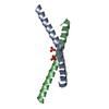
| ||||||||||||
|---|---|---|---|---|---|---|---|---|---|---|---|---|---|
| 1 | 
| ||||||||||||
| Unit cell |
|
-Components
| #1: Protein/peptide | Mass: 5184.021 Da / Num. of mol.: 2 Source method: isolated from a genetically manipulated source Source: (gene. exp.) Homo sapiens (human) / Gene: AAGAB / Production host:  #2: Chemical |   Mass: 94.971 Da / Num. of mol.: 2 / Source method: obtained synthetically / Formula: PO4 Mass: 94.971 Da / Num. of mol.: 2 / Source method: obtained synthetically / Formula: PO4#3: Water | ChemComp-HOH / |  Mass: 18.015 Da / Num. of mol.: 74 / Source method: isolated from a natural source / Formula: H2O Mass: 18.015 Da / Num. of mol.: 74 / Source method: isolated from a natural source / Formula: H2OHas ligand of interest | N | |
|---|
-Experimental details
-Experiment
| Experiment | Method: X-RAY DIFFRACTION / Number of used crystals: 1 |
|---|
- Sample preparation
| Crystal | Density Matthews: 3.01 Å3/Da / Density % sol: 59.15 % |
|---|---|
| Crystal grow | Temperature: 289 K / Method: vapor diffusion, hanging drop / pH: 7.5 / Details: 30% glycerol, 0.5 M ammonium phosphate |
-Data collection
| Diffraction | Mean temperature: 100 K / Serial crystal experiment: N |
|---|---|
| Diffraction source | Source: SYNCHROTRON / Site: APS / Beamline: 22-ID / Wavelength: 1 Å |
| Detector | Type: DECTRIS EIGER X 16M / Detector: PIXEL / Date: Nov 7, 2018 |
| Radiation | Protocol: SINGLE WAVELENGTH / Monochromatic (M) / Laue (L): M / Scattering type: x-ray |
| Radiation wavelength | Wavelength: 1 Å / Relative weight: 1 |
| Reflection | Resolution: 2→50 Å / Num. obs: 9351 / % possible obs: 99.2 % / Redundancy: 13.5 % / Biso Wilson estimate: 22.72 Å2 / CC1/2: 0.985 / CC star: 0.996 / Rmerge(I) obs: 0.205 / Rpim(I) all: 0.057 / Rrim(I) all: 0.213 / Χ2: 0.458 / Net I/σ(I): 17.4 |
| Reflection shell | Resolution: 2→2.07 Å / Redundancy: 9.6 % / Rmerge(I) obs: 1.58 / Mean I/σ(I) obs: 0.929 / Num. unique obs: 900 / CC1/2: 0.38 / CC star: 0.742 / Rpim(I) all: 0.479 / Rrim(I) all: 1.658 / Χ2: 0.458 / % possible all: 99.7 |
- Processing
| Software |
| |||||||||||||||||||||||||||||||||||||||||||||||||
|---|---|---|---|---|---|---|---|---|---|---|---|---|---|---|---|---|---|---|---|---|---|---|---|---|---|---|---|---|---|---|---|---|---|---|---|---|---|---|---|---|---|---|---|---|---|---|---|---|---|---|
| Refinement | Method to determine structure: SAD / Resolution: 2.11→40.25 Å / SU ML: 0.2164 / Cross valid method: FREE R-VALUE / σ(F): 1.38 / Phase error: 23.4306 Stereochemistry target values: GeoStd + Monomer Library + CDL v1.2
| |||||||||||||||||||||||||||||||||||||||||||||||||
| Solvent computation | Shrinkage radii: 0.9 Å / VDW probe radii: 1.11 Å / Solvent model: FLAT BULK SOLVENT MODEL | |||||||||||||||||||||||||||||||||||||||||||||||||
| Displacement parameters | Biso mean: 35.63 Å2 | |||||||||||||||||||||||||||||||||||||||||||||||||
| Refinement step | Cycle: LAST / Resolution: 2.11→40.25 Å
| |||||||||||||||||||||||||||||||||||||||||||||||||
| Refine LS restraints |
| |||||||||||||||||||||||||||||||||||||||||||||||||
| LS refinement shell |
| |||||||||||||||||||||||||||||||||||||||||||||||||
| Refinement TLS params. | Method: refined / Origin x: -24.1235768997 Å / Origin y: 10.4592267274 Å / Origin z: -1.3874935756 Å
| |||||||||||||||||||||||||||||||||||||||||||||||||
| Refinement TLS group | Selection details: all |
