 Movie
Movie Controller

[English] 日本語
 Yorodumi
- PDB-7ttv: E.coli DsbA in complex with 4-phenyl-2-(3-phenylpropyl)thiazole-5... -
+ Open data

- Basic information
| Entry | Database: PDB / ID: 7ttv | ||||||
|---|---|---|---|---|---|---|---|
| Title | E.coli DsbA in complex with 4-phenyl-2-(3-phenylpropyl)thiazole-5-carboxylic acid | ||||||
 Components Components | Thiol:disulfide interchange protein DsbA | ||||||
 Keywords Keywords | OXIDOREDUCTASE/OXIDOREDUCTASE INHIBITOR / Inhibitor / complex / disulfide oxidoreductase / fragments / OXIDOREDUCTASE / OXIDOREDUCTASE-OXIDOREDUCTASE INHIBITOR complex | ||||||
| Function / homology |  Function and homology information Function and homology informationcellular response to antibiotic / protein disulfide isomerase activity / protein-disulfide reductase activity / outer membrane-bounded periplasmic space Similarity search - Function | ||||||
| Biological species |  | ||||||
| Method |  X-RAY DIFFRACTION / SYNCHROTRON / MOLECULAR REPLACEMENT / Resolution: 1.99 Å X-RAY DIFFRACTION / SYNCHROTRON / MOLECULAR REPLACEMENT / Resolution: 1.99 Å | ||||||
 Authors Authors | Wang, G. / Heras, B. | ||||||
| Funding support |  Australia, 1items Australia, 1items
| ||||||
 Citation Citation | Journal: Sci Rep / Year: 2022 Title: Methyl probes in proteins for determining ligand binding mode in weak protein-ligand complexes. Authors: Mohanty, B. / Orts, J. / Wang, G. / Nebl, S. / Alwan, W.S. / Doak, B.C. / Williams, M.L. / Heras, B. / Mobli, M. / Scanlon, M.J. | ||||||
| History |
|
- Structure visualization
| Structure viewer | Molecule: MolmilJmol/JSmol |
|---|
- Downloads & links
-Download
| PDBx/mmCIF format | 7ttv.cif.gz | 97.5 KB | Display | PDBx/mmCIF format |
|---|---|---|---|---|
| PDB format | pdb7ttv.ent.gz | 72 KB | Display | PDB format |
| PDBx/mmJSON format | 7ttv.json.gz | Tree view | PDBx/mmJSON format | |
| Others |  Other downloads Other downloads |
-Validation report
| Summary document | 7ttv_validation.pdf.gz | 738.8 KB | Display | wwPDB validaton report |
|---|---|---|---|---|
| Full document | 7ttv_full_validation.pdf.gz | 743.1 KB | Display | |
| Data in XML | 7ttv_validation.xml.gz | 19.7 KB | Display | |
| Data in CIF | 7ttv_validation.cif.gz | 29 KB | Display | |
| Arichive directory | https://data.pdbj.org/pub/pdb/validation_reports/tt/7ttvftp://data.pdbj.org/pub/pdb/validation_reports/tt/7ttv | HTTPS FTP |
-Related structure data
| Related structure data |  1fvkS S: Starting model for refinement |
|---|---|
| Similar structure data |
-Links
 PDBj
PDBj

- Assembly
| Deposited unit | 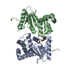
| ||||||||
|---|---|---|---|---|---|---|---|---|---|
| 1 | 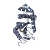
| ||||||||
| 2 | 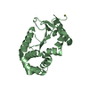
| ||||||||
| Unit cell |
| ||||||||
| Components on special symmetry positions |
|
-Components
| #1: Protein | Mass: 21155.025 Da / Num. of mol.: 2 Source method: isolated from a genetically manipulated source Source: (gene. exp.) #2: Chemical | ChemComp-QVP / | 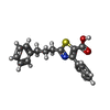  Mass: 323.409 Da / Num. of mol.: 1 / Source method: obtained synthetically / Formula: C19H17NO2S / Feature type: SUBJECT OF INVESTIGATION Mass: 323.409 Da / Num. of mol.: 1 / Source method: obtained synthetically / Formula: C19H17NO2S / Feature type: SUBJECT OF INVESTIGATION#3: Chemical | ChemComp-CU / |   Mass: 63.546 Da / Num. of mol.: 1 / Source method: obtained synthetically / Formula: Cu Mass: 63.546 Da / Num. of mol.: 1 / Source method: obtained synthetically / Formula: Cu#4: Water | ChemComp-HOH / |  Mass: 18.015 Da / Num. of mol.: 349 / Source method: isolated from a natural source / Formula: H2O Mass: 18.015 Da / Num. of mol.: 349 / Source method: isolated from a natural source / Formula: H2OHas ligand of interest | Y | Has protein modification | Y | |
|---|
-Experimental details
-Experiment
| Experiment | Method: X-RAY DIFFRACTION / Number of used crystals: 1 |
|---|
- Sample preparation
| Crystal | Density Matthews: 2.66 Å3/Da / Density % sol: 53.8 % |
|---|---|
| Crystal grow | Temperature: 293 K / Method: vapor diffusion Details: 11-13% PEG 8000, 5-7.5% glycerol, 1 mM copper(II) chloride, 100 mM sodium cacodylate |
-Data collection
| Diffraction | Mean temperature: 100 K / Serial crystal experiment: N | |||||||||||||||||||||||||||||||||||||||||||||||||||||||||||||||||||||||||||||||||||||||||||||||||||||||||||||||||||||||||
|---|---|---|---|---|---|---|---|---|---|---|---|---|---|---|---|---|---|---|---|---|---|---|---|---|---|---|---|---|---|---|---|---|---|---|---|---|---|---|---|---|---|---|---|---|---|---|---|---|---|---|---|---|---|---|---|---|---|---|---|---|---|---|---|---|---|---|---|---|---|---|---|---|---|---|---|---|---|---|---|---|---|---|---|---|---|---|---|---|---|---|---|---|---|---|---|---|---|---|---|---|---|---|---|---|---|---|---|---|---|---|---|---|---|---|---|---|---|---|---|---|---|---|
| Diffraction source | Source: SYNCHROTRON / Site: Australian Synchrotron / Beamline: MX1 / Wavelength: 0.9537 Å | |||||||||||||||||||||||||||||||||||||||||||||||||||||||||||||||||||||||||||||||||||||||||||||||||||||||||||||||||||||||||
| Detector | Type: ADSC QUANTUM 210r / Detector: CCD / Date: Aug 3, 2016 | |||||||||||||||||||||||||||||||||||||||||||||||||||||||||||||||||||||||||||||||||||||||||||||||||||||||||||||||||||||||||
| Radiation | Protocol: SINGLE WAVELENGTH / Monochromatic (M) / Laue (L): M / Scattering type: x-ray | |||||||||||||||||||||||||||||||||||||||||||||||||||||||||||||||||||||||||||||||||||||||||||||||||||||||||||||||||||||||||
| Radiation wavelength | Wavelength: 0.9537 Å / Relative weight: 1 | |||||||||||||||||||||||||||||||||||||||||||||||||||||||||||||||||||||||||||||||||||||||||||||||||||||||||||||||||||||||||
| Reflection | Resolution: 1.986→60.193 Å / Num. all: 30753 / Num. obs: 30753 / % possible obs: 99.7 % / Redundancy: 4.1 % / Biso Wilson estimate: 23.88 Å2 / Rpim(I) all: 0.05 / Rrim(I) all: 0.101 / Rsym value: 0.088 / Net I/av σ(I): 7.9 / Net I/σ(I): 11.5 / Num. measured all: 125514 | |||||||||||||||||||||||||||||||||||||||||||||||||||||||||||||||||||||||||||||||||||||||||||||||||||||||||||||||||||||||||
| Reflection shell | Diffraction-ID: 1
|
- Processing
| Software |
| |||||||||||||||||||||||||||||||||||||||||||||||||||||||||||||||||||||||||||||
|---|---|---|---|---|---|---|---|---|---|---|---|---|---|---|---|---|---|---|---|---|---|---|---|---|---|---|---|---|---|---|---|---|---|---|---|---|---|---|---|---|---|---|---|---|---|---|---|---|---|---|---|---|---|---|---|---|---|---|---|---|---|---|---|---|---|---|---|---|---|---|---|---|---|---|---|---|---|---|
| Refinement | Method to determine structure: MOLECULAR REPLACEMENT Starting model: 1FVK Resolution: 1.99→37.08 Å / SU ML: 0.25 / Cross valid method: THROUGHOUT / σ(F): 1.34 / Phase error: 23.91 / Stereochemistry target values: ML
| |||||||||||||||||||||||||||||||||||||||||||||||||||||||||||||||||||||||||||||
| Solvent computation | Shrinkage radii: 0.9 Å / VDW probe radii: 1.11 Å / Solvent model: FLAT BULK SOLVENT MODEL | |||||||||||||||||||||||||||||||||||||||||||||||||||||||||||||||||||||||||||||
| Displacement parameters | Biso max: 111.64 Å2 / Biso mean: 29.0546 Å2 / Biso min: 10.83 Å2 | |||||||||||||||||||||||||||||||||||||||||||||||||||||||||||||||||||||||||||||
| Refinement step | Cycle: final / Resolution: 1.99→37.08 Å
| |||||||||||||||||||||||||||||||||||||||||||||||||||||||||||||||||||||||||||||
| LS refinement shell | Refine-ID: X-RAY DIFFRACTION / Rfactor Rfree error: 0 / Total num. of bins used: 10
|
