 Movie
Movie Controller

[English] 日本語
 Yorodumi
- PDB-7tsj: Xenon-bound structure of carbon monoxide dehydrogenase (CODH) fro... -
+ Open data

- Basic information
| Entry | Database: PDB / ID: 7tsj | ||||||||||||
|---|---|---|---|---|---|---|---|---|---|---|---|---|---|
| Title | Xenon-bound structure of carbon monoxide dehydrogenase (CODH) from Desulfovibrio vulgaris | ||||||||||||
 Components Components | Carbon monoxide dehydrogenase | ||||||||||||
 Keywords Keywords | OXIDOREDUCTASE / CO-dehydrogenase / Desulfovibrio vulgaris / Xenon | ||||||||||||
| Function / homology |  Function and homology information Function and homology informationanaerobic carbon monoxide dehydrogenase / anaerobic carbon-monoxide dehydrogenase activity / : / hydroxylamine reductase activity / nickel cation binding / generation of precursor metabolites and energy / peroxidase activity / response to hydrogen peroxide / 2 iron, 2 sulfur cluster binding / 4 iron, 4 sulfur cluster binding Similarity search - Function | ||||||||||||
| Biological species |  Desulfovibrio vulgaris (bacteria) Desulfovibrio vulgaris (bacteria) | ||||||||||||
| Method |  X-RAY DIFFRACTION / SYNCHROTRON / MOLECULAR REPLACEMENT / Resolution: 2.1 Å X-RAY DIFFRACTION / SYNCHROTRON / MOLECULAR REPLACEMENT / Resolution: 2.1 Å | ||||||||||||
 Authors Authors | Biester, A. / Drennan, C.L. | ||||||||||||
| Funding support |  United States, United States,  Canada, 3items Canada, 3items
| ||||||||||||
 Citation Citation | Journal: J.Inorg.Biochem. / Year: 2022 Title: Visualizing the gas channel of a monofunctional carbon monoxide dehydrogenase. Authors: Biester, A. / Dementin, S. / Drennan, C.L. | ||||||||||||
| History |
|
- Structure visualization
| Structure viewer | Molecule: MolmilJmol/JSmol |
|---|
- Downloads & links
-Download
| PDBx/mmCIF format | 7tsj.cif.gz | 250.9 KB | Display | PDBx/mmCIF format |
|---|---|---|---|---|
| PDB format | pdb7tsj.ent.gz | 198 KB | Display | PDB format |
| PDBx/mmJSON format | 7tsj.json.gz | Tree view | PDBx/mmJSON format | |
| Others |  Other downloads Other downloads |
-Validation report
| Arichive directory | https://data.pdbj.org/pub/pdb/validation_reports/ts/7tsjftp://data.pdbj.org/pub/pdb/validation_reports/ts/7tsj | HTTPS FTP |
|---|
-Related structure data
| Related structure data |  6b6vS S: Starting model for refinement |
|---|---|
| Similar structure data |
-Links
 PDBj
PDBj


- Assembly
| Deposited unit | 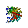
| ||||||||
|---|---|---|---|---|---|---|---|---|---|
| 1 | 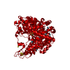
| ||||||||
| Unit cell |
|
-Components
-Protein , 1 types, 1 molecules A
| #1: Protein | Mass: 67715.898 Da / Num. of mol.: 1 Source method: isolated from a genetically manipulated source Source: (gene. exp.) Desulfovibrio vulgaris (bacteria) / Gene: cooS, DVU_2098Production host: Solidesulfovibrio fructosivorans (bacteria)References: UniProt: Q72A99, anaerobic carbon monoxide dehydrogenase |
|---|
-Non-polymers , 5 types, 89 molecules 


| #2: Chemical | ChemComp-SF4 /  Mass: 351.640 Da / Num. of mol.: 1 / Source method: isolated from a natural source / Formula: Fe4S4 Mass: 351.640 Da / Num. of mol.: 1 / Source method: isolated from a natural source / Formula: Fe4S4 | ||
|---|---|---|---|
| #3: Chemical | ChemComp-FES /  Mass: 175.820 Da / Num. of mol.: 1 / Source method: isolated from a natural source / Formula: Fe2S2 Mass: 175.820 Da / Num. of mol.: 1 / Source method: isolated from a natural source / Formula: Fe2S2 | ||
| #4: Chemical | ChemComp-CUV /  Mass: 410.333 Da / Num. of mol.: 1 / Source method: isolated from a natural source / Formula: Fe4NiS4 Mass: 410.333 Da / Num. of mol.: 1 / Source method: isolated from a natural source / Formula: Fe4NiS4 | ||
| #5: Chemical | ChemComp-XE /  Mass: 131.293 Da / Num. of mol.: 12 / Source method: obtained synthetically / Formula: Xe / Feature type: SUBJECT OF INVESTIGATION Mass: 131.293 Da / Num. of mol.: 12 / Source method: obtained synthetically / Formula: Xe / Feature type: SUBJECT OF INVESTIGATION#6: Water | ChemComp-HOH / | Mass: 18.015 Da / Num. of mol.: 74 / Source method: isolated from a natural source / Formula: H2O |
-Details
| Has ligand of interest | Y |
|---|
-Experimental details
-Experiment
| Experiment | Method: X-RAY DIFFRACTION / Number of used crystals: 1 |
|---|
- Sample preparation
| Crystal | Density Matthews: 2.3 Å3/Da / Density % sol: 46.6 % |
|---|---|
| Crystal grow | Temperature: 298 K / Method: vapor diffusion, hanging drop / pH: 8 / Details: 250 mM MgCl2, 16% (w/v) PEG 3350 |
-Data collection
| Diffraction | Mean temperature: 100 K / Serial crystal experiment: N | ||||||||||||||||||||||||||||||||||||||||||||||||||||||||||||||||||||||||||||||||||||||||||||||||||||
|---|---|---|---|---|---|---|---|---|---|---|---|---|---|---|---|---|---|---|---|---|---|---|---|---|---|---|---|---|---|---|---|---|---|---|---|---|---|---|---|---|---|---|---|---|---|---|---|---|---|---|---|---|---|---|---|---|---|---|---|---|---|---|---|---|---|---|---|---|---|---|---|---|---|---|---|---|---|---|---|---|---|---|---|---|---|---|---|---|---|---|---|---|---|---|---|---|---|---|---|---|---|
| Diffraction source | Source: SYNCHROTRON / Site: APS / Beamline: 24-ID-C / Wavelength: 0.9791 Å | ||||||||||||||||||||||||||||||||||||||||||||||||||||||||||||||||||||||||||||||||||||||||||||||||||||
| Detector | Type: DECTRIS EIGER2 X 16M / Detector: PIXEL / Date: Dec 2, 2020 | ||||||||||||||||||||||||||||||||||||||||||||||||||||||||||||||||||||||||||||||||||||||||||||||||||||
| Radiation | Protocol: SINGLE WAVELENGTH / Monochromatic (M) / Laue (L): M / Scattering type: x-ray | ||||||||||||||||||||||||||||||||||||||||||||||||||||||||||||||||||||||||||||||||||||||||||||||||||||
| Radiation wavelength | Wavelength: 0.9791 Å / Relative weight: 1 | ||||||||||||||||||||||||||||||||||||||||||||||||||||||||||||||||||||||||||||||||||||||||||||||||||||
| Reflection | Resolution: 2.1→46.72 Å / Num. obs: 34865 / % possible obs: 97.7 % / Redundancy: 3.867 % / Biso Wilson estimate: 40.8 Å2 / CC1/2: 0.998 / Rmerge(I) obs: 0.062 / Rrim(I) all: 0.071 / Χ2: 0.786 / Net I/σ(I): 12.57 / Num. measured all: 134825 | ||||||||||||||||||||||||||||||||||||||||||||||||||||||||||||||||||||||||||||||||||||||||||||||||||||
| Reflection shell | Diffraction-ID: 1
|
- Processing
| Software |
| ||||||||||||||||||||||||||||||||||||||||||||||||||||||||||||||||||||||||||||||||||||||||||||||||||
|---|---|---|---|---|---|---|---|---|---|---|---|---|---|---|---|---|---|---|---|---|---|---|---|---|---|---|---|---|---|---|---|---|---|---|---|---|---|---|---|---|---|---|---|---|---|---|---|---|---|---|---|---|---|---|---|---|---|---|---|---|---|---|---|---|---|---|---|---|---|---|---|---|---|---|---|---|---|---|---|---|---|---|---|---|---|---|---|---|---|---|---|---|---|---|---|---|---|---|---|
| Refinement | Method to determine structure: MOLECULAR REPLACEMENT Starting model: 6B6V Resolution: 2.1→46.72 Å / SU ML: 0.25 / Cross valid method: THROUGHOUT / σ(F): 1.39 / Phase error: 22.7 / Stereochemistry target values: ML
| ||||||||||||||||||||||||||||||||||||||||||||||||||||||||||||||||||||||||||||||||||||||||||||||||||
| Solvent computation | Shrinkage radii: 0.9 Å / VDW probe radii: 1.1 Å / Solvent model: FLAT BULK SOLVENT MODEL | ||||||||||||||||||||||||||||||||||||||||||||||||||||||||||||||||||||||||||||||||||||||||||||||||||
| Displacement parameters | Biso max: 115.93 Å2 / Biso mean: 52.9908 Å2 / Biso min: 27.87 Å2 | ||||||||||||||||||||||||||||||||||||||||||||||||||||||||||||||||||||||||||||||||||||||||||||||||||
| Refinement step | Cycle: final / Resolution: 2.1→46.72 Å
| ||||||||||||||||||||||||||||||||||||||||||||||||||||||||||||||||||||||||||||||||||||||||||||||||||
| LS refinement shell | Refine-ID: X-RAY DIFFRACTION / Rfactor Rfree error: 0 / Total num. of bins used: 13
| ||||||||||||||||||||||||||||||||||||||||||||||||||||||||||||||||||||||||||||||||||||||||||||||||||
| Refinement TLS params. | Method: refined / Origin x: 19.232 Å / Origin y: -5.7906 Å / Origin z: 1.5046 Å
| ||||||||||||||||||||||||||||||||||||||||||||||||||||||||||||||||||||||||||||||||||||||||||||||||||
| Refinement TLS group |
|
