 Movie
Movie Controller

[English] 日本語
 Yorodumi
- PDB-7tpm: Structure of the outer-membrane lipoprotein carrier protein (LolA... -
+ Open data

- Basic information
| Entry | Database: PDB / ID: 7tpm | ||||||
|---|---|---|---|---|---|---|---|
| Title | Structure of the outer-membrane lipoprotein carrier protein (LolA) from Borrelia burgdorferi | ||||||
 Components Components | Outer membrane lipoprotein carrier protein LolA | ||||||
 Keywords Keywords | LIPID TRANSPORT / lipoprotein carrier protein / LolA / Borreliella burgdorferi / Lyme disease spirochete | ||||||
| Function / homology | Outer membrane lipoprotein carrier protein LolA / Outer membrane lipoprotein carrier protein LolA-like / Lipoprotein localisation LolA/LolB/LppX / Export chaperone Function and homology information Function and homology information | ||||||
| Biological species |  Borreliella burgdorferi (Lyme disease spirochete) Borreliella burgdorferi (Lyme disease spirochete) | ||||||
| Method |  X-RAY DIFFRACTION / SYNCHROTRON / MOLECULAR REPLACEMENT / SAD / Resolution: 1.9 Å X-RAY DIFFRACTION / SYNCHROTRON / MOLECULAR REPLACEMENT / SAD / Resolution: 1.9 Å | ||||||
 Authors Authors | Lovell, S. / Kashipathy, M.M. / Battaile, K.P. / Zueckert, W.R. | ||||||
| Funding support |  United States, 1items United States, 1items
| ||||||
 Citation Citation | Journal: To be published Title: Structure of the outer-membrane lipoprotein carrier protein (LolA) from Borrelia burgdorferi Authors: Lovell, S. / Kashipathy, M.M. / Battaile, K.P. / Zueckert, W.R. | ||||||
| History |
|
- Structure visualization
| Structure viewer | Molecule: MolmilJmol/JSmol |
|---|
- Downloads & links
-Download
| PDBx/mmCIF format | 7tpm.cif.gz | 57.5 KB | Display | PDBx/mmCIF format |
|---|---|---|---|---|
| PDB format | pdb7tpm.ent.gz | 39.8 KB | Display | PDB format |
| PDBx/mmJSON format | 7tpm.json.gz | Tree view | PDBx/mmJSON format | |
| Others |  Other downloads Other downloads |
-Validation report
| Summary document | 7tpm_validation.pdf.gz | 682.1 KB | Display | wwPDB validaton report |
|---|---|---|---|---|
| Full document | 7tpm_full_validation.pdf.gz | 683 KB | Display | |
| Data in XML | 7tpm_validation.xml.gz | 11.2 KB | Display | |
| Data in CIF | 7tpm_validation.cif.gz | 15.6 KB | Display | |
| Arichive directory | https://data.pdbj.org/pub/pdb/validation_reports/tp/7tpmftp://data.pdbj.org/pub/pdb/validation_reports/tp/7tpm | HTTPS FTP |
-Related structure data
| Similar structure data |
|---|
-Links
 PDBj
PDBj- Assembly
| Deposited unit | 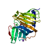
| ||||||||
|---|---|---|---|---|---|---|---|---|---|
| 1 |
| ||||||||
| Unit cell |
|
-Components
| #1: Protein | Mass: 23403.543 Da / Num. of mol.: 1 / Fragment: Q18-N216 Source method: isolated from a genetically manipulated source Source: (gene. exp.) Borreliella burgdorferi (Lyme disease spirochete)Gene: BB_0346 / Plasmid: pET29b / Production host: | ||||||
|---|---|---|---|---|---|---|---|
| #2: Chemical | ChemComp-PE4 /   Mass: 354.436 Da / Num. of mol.: 1 / Source method: obtained synthetically / Formula: C16H34O8 / Feature type: SUBJECT OF INVESTIGATION / Comment: precipitant*YM Mass: 354.436 Da / Num. of mol.: 1 / Source method: obtained synthetically / Formula: C16H34O8 / Feature type: SUBJECT OF INVESTIGATION / Comment: precipitant*YM | ||||||
| #3: Chemical |   Mass: 35.453 Da / Num. of mol.: 3 / Source method: obtained synthetically / Formula: Cl Mass: 35.453 Da / Num. of mol.: 3 / Source method: obtained synthetically / Formula: Cl#4: Chemical | ChemComp-NA / |   Mass: 22.990 Da / Num. of mol.: 1 / Source method: obtained synthetically / Formula: Na Mass: 22.990 Da / Num. of mol.: 1 / Source method: obtained synthetically / Formula: Na#5: Water | ChemComp-HOH / |  Mass: 18.015 Da / Num. of mol.: 158 / Source method: isolated from a natural source / Formula: H2O Mass: 18.015 Da / Num. of mol.: 158 / Source method: isolated from a natural source / Formula: H2OHas ligand of interest | Y | |
-Experimental details
-Experiment
| Experiment | Method: X-RAY DIFFRACTION / Number of used crystals: 1 |
|---|
- Sample preparation
| Crystal | Density Matthews: 3.24 Å3/Da / Density % sol: 62.08 % / Mosaicity: 0.04 ° |
|---|---|
| Crystal grow | Temperature: 291 K / Method: vapor diffusion, sitting drop / pH: 5.5 Details: 2 M sodium formate, 100 mM MES, 5% (w/v) PEG 5000 MME |
-Data collection
| Diffraction | Mean temperature: 100 K / Serial crystal experiment: N | ||||||||||||||||||||||||
|---|---|---|---|---|---|---|---|---|---|---|---|---|---|---|---|---|---|---|---|---|---|---|---|---|---|
| Diffraction source | Source: SYNCHROTRON / Site: APS / Beamline: 17-ID / Wavelength: 1 Å | ||||||||||||||||||||||||
| Detector | Type: DECTRIS PILATUS 6M / Detector: PIXEL / Date: Mar 4, 2018 | ||||||||||||||||||||||||
| Radiation | Protocol: SINGLE WAVELENGTH / Monochromatic (M) / Laue (L): M / Scattering type: x-ray | ||||||||||||||||||||||||
| Radiation wavelength | Wavelength: 1 Å / Relative weight: 1 | ||||||||||||||||||||||||
| Reflection | Resolution: 1.9→49.86 Å / Num. obs: 25182 / % possible obs: 100 % / Redundancy: 19.4 % / Biso Wilson estimate: 33.39 Å2 / CC1/2: 0.999 / Rmerge(I) obs: 0.145 / Net I/σ(I): 12.5 / Num. measured all: 488064 / Scaling rejects: 1 | ||||||||||||||||||||||||
| Reflection shell | Diffraction-ID: 1
|
-Phasing
| Phasing | Method: SAD |
|---|
- Processing
| Software |
| ||||||||||||||||||||||||||||||||||||||||||||||||||
|---|---|---|---|---|---|---|---|---|---|---|---|---|---|---|---|---|---|---|---|---|---|---|---|---|---|---|---|---|---|---|---|---|---|---|---|---|---|---|---|---|---|---|---|---|---|---|---|---|---|---|---|
| Refinement | Method to determine structure: MOLECULAR REPLACEMENT Starting model: SAD-phased structure of the same protein from a KI-soaked crystal Resolution: 1.9→38.623 Å / SU ML: 0.21 / Cross valid method: THROUGHOUT / σ(F): 1.06 / Phase error: 24.52 / Stereochemistry target values: ML
| ||||||||||||||||||||||||||||||||||||||||||||||||||
| Solvent computation | Shrinkage radii: 0.9 Å / VDW probe radii: 1.11 Å / Solvent model: FLAT BULK SOLVENT MODEL | ||||||||||||||||||||||||||||||||||||||||||||||||||
| Displacement parameters | Biso max: 112.69 Å2 / Biso mean: 37.2703 Å2 / Biso min: 20.3 Å2 | ||||||||||||||||||||||||||||||||||||||||||||||||||
| Refinement step | Cycle: final / Resolution: 1.9→38.623 Å
| ||||||||||||||||||||||||||||||||||||||||||||||||||
| Refine LS restraints |
| ||||||||||||||||||||||||||||||||||||||||||||||||||
| LS refinement shell | Refine-ID: X-RAY DIFFRACTION / Rfactor Rfree error: 0 / % reflection obs: 100 %
|
