 Movie
Movie Controller

[English] 日本語
 Yorodumi
- PDB-7tmh: Porous framework formed by assembly of a bipyridyl-conjugated hel... -
+ Open data

- Basic information
| Entry | Database: PDB / ID: 7tmh | |||||||||
|---|---|---|---|---|---|---|---|---|---|---|
| Title | Porous framework formed by assembly of a bipyridyl-conjugated helical peptide | |||||||||
 Components Components | bipyridyl-conjugated helical peptide | |||||||||
 Keywords Keywords | DE NOVO PROTEIN / porous / framework / helix / 310 / alpha / assembly / bipyridine / UIC-1 | |||||||||
| Function / homology | ACETONITRILE Function and homology information Function and homology information | |||||||||
| Biological species | synthetic construct (others) | |||||||||
| Method |  X-RAY DIFFRACTION / SYNCHROTRON / MOLECULAR REPLACEMENT / Resolution: 0.8 Å X-RAY DIFFRACTION / SYNCHROTRON / MOLECULAR REPLACEMENT / Resolution: 0.8 Å | |||||||||
 Authors Authors | Nguyen, A.I. | |||||||||
| Funding support | 1items
| |||||||||
 Citation Citation | Journal: J.Am.Chem.Soc. / Year: 2022 Title: Assembly of pi-Stacking Helical Peptides into a Porous and Multivariable Proteomimetic Framework. Authors: Heinz-Kunert, S.L. / Pandya, A. / Dang, V.T. / Tran, P.N. / Ghosh, S. / McElheny, D. / Santarsiero, B.D. / Ren, Z. / Nguyen, A.I. | |||||||||
| History |
|
- Structure visualization
| Structure viewer | Molecule: MolmilJmol/JSmol |
|---|
- Downloads & links
-Download
| PDBx/mmCIF format | 7tmh.cif.gz | 25 KB | Display | PDBx/mmCIF format |
|---|---|---|---|---|
| PDB format | pdb7tmh.ent.gz | 13.9 KB | Display | PDB format |
| PDBx/mmJSON format | 7tmh.json.gz | Tree view | PDBx/mmJSON format | |
| Others |  Other downloads Other downloads |
-Validation report
| Arichive directory | https://data.pdbj.org/pub/pdb/validation_reports/tm/7tmhftp://data.pdbj.org/pub/pdb/validation_reports/tm/7tmh | HTTPS FTP |
|---|
-Related structure data
| Related structure data | 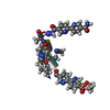 7tlsSC 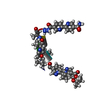 7tluC 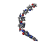 7tm1C 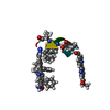 7tm2C 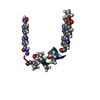 7tmaC 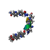 7tmeC  7tmiC 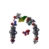 7tmjC  7tmkC 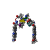 7tmlC S: Starting model for refinement C: citing same article ( |
|---|---|
| Similar structure data |
-Links
 PDBj
PDBj
- Assembly
| Deposited unit | 
| ||||||||||||
|---|---|---|---|---|---|---|---|---|---|---|---|---|---|
| 1 |
| ||||||||||||
| Unit cell |
|
-Components
| #1: Protein/peptide | Mass: 1624.646 Da / Num. of mol.: 1 / Source method: obtained synthetically / Source: (synth.) synthetic construct (others) | ||||
|---|---|---|---|---|---|
| #2: Chemical |   Mass: 41.052 Da / Num. of mol.: 2 / Source method: obtained synthetically / Formula: C2H3N Mass: 41.052 Da / Num. of mol.: 2 / Source method: obtained synthetically / Formula: C2H3N#3: Water | ChemComp-HOH / |  Mass: 18.015 Da / Num. of mol.: 5 / Source method: isolated from a natural source / Formula: H2O Mass: 18.015 Da / Num. of mol.: 5 / Source method: isolated from a natural source / Formula: H2OHas ligand of interest | N | |
-Experimental details
-Experiment
| Experiment | Method: X-RAY DIFFRACTION / Number of used crystals: 1 |
|---|
- Sample preparation
| Crystal | Density Matthews: 2.18 Å3/Da / Density % sol: 43.61 % |
|---|---|
| Crystal grow | Temperature: 295 K / Method: slow cooling / Details: water and acetonitrile |
-Data collection
| Diffraction | Mean temperature: 100 K / Serial crystal experiment: N |
|---|---|
| Diffraction source | Source: SYNCHROTRON / Site: APS  / Beamline: 21-ID-D / Wavelength: 0.688 Å / Beamline: 21-ID-D / Wavelength: 0.688 Å |
| Detector | Type: DECTRIS EIGER X 9M / Detector: PIXEL / Date: Jul 1, 2021 |
| Radiation | Protocol: SINGLE WAVELENGTH / Monochromatic (M) / Laue (L): M / Scattering type: x-ray |
| Radiation wavelength | Wavelength: 0.688 Å / Relative weight: 1 |
| Reflection | Resolution: 0.8→13.5 Å / Num. obs: 19340 / % possible obs: 95.43 % / Redundancy: 6.7 % / Biso Wilson estimate: 4.95 Å2 / Rrim(I) all: 0.05818 / Net I/σ(I): 26.34 |
| Reflection shell | Resolution: 0.83→0.8597 Å / Num. unique obs: 1826 / Rrim(I) all: 0.1462 |
- Processing
| Software |
| |||||||||||||||||||||||||||||||||||||||||||||||||||||||||||||||||||||||||||||||||||||||||||||||||||||||||
|---|---|---|---|---|---|---|---|---|---|---|---|---|---|---|---|---|---|---|---|---|---|---|---|---|---|---|---|---|---|---|---|---|---|---|---|---|---|---|---|---|---|---|---|---|---|---|---|---|---|---|---|---|---|---|---|---|---|---|---|---|---|---|---|---|---|---|---|---|---|---|---|---|---|---|---|---|---|---|---|---|---|---|---|---|---|---|---|---|---|---|---|---|---|---|---|---|---|---|---|---|---|---|---|---|---|---|
| Refinement | Method to determine structure: MOLECULAR REPLACEMENT Starting model: 7TLS Resolution: 0.8→13.5 Å / SU ML: 0.0456 / Cross valid method: FREE R-VALUE / σ(F): 1.4 / Phase error: 13.0185 Stereochemistry target values: GeoStd + Monomer Library + CDL v1.2
| |||||||||||||||||||||||||||||||||||||||||||||||||||||||||||||||||||||||||||||||||||||||||||||||||||||||||
| Solvent computation | Shrinkage radii: 0.9 Å / VDW probe radii: 1.11 Å / Solvent model: FLAT BULK SOLVENT MODEL | |||||||||||||||||||||||||||||||||||||||||||||||||||||||||||||||||||||||||||||||||||||||||||||||||||||||||
| Displacement parameters | Biso mean: 6.85 Å2 | |||||||||||||||||||||||||||||||||||||||||||||||||||||||||||||||||||||||||||||||||||||||||||||||||||||||||
| Refinement step | Cycle: LAST / Resolution: 0.8→13.5 Å
| |||||||||||||||||||||||||||||||||||||||||||||||||||||||||||||||||||||||||||||||||||||||||||||||||||||||||
| Refine LS restraints |
| |||||||||||||||||||||||||||||||||||||||||||||||||||||||||||||||||||||||||||||||||||||||||||||||||||||||||
| LS refinement shell |
|
