 Movie
Movie Controller


+ Open data

- Basic information
| Entry | Database: PDB / ID: 7tlj | ||||||
|---|---|---|---|---|---|---|---|
| Title | Rhodobacter sphaeroides Mitochondrial respiratory chain complex | ||||||
 Components Components |
| ||||||
 Keywords Keywords | OXIDOREDUCTASE / Mitochondrial respiratory chain complex / cytochrome bc1 / inhibitors / electron transfer | ||||||
| Function / homology |  Function and homology information Function and homology informationrespiratory chain complex III / quinol-cytochrome-c reductase / quinol-cytochrome-c reductase activity / respiratory electron transport chain / 2 iron, 2 sulfur cluster binding / electron transfer activity / oxidoreductase activity / heme binding / metal ion binding / plasma membrane Similarity search - Function | ||||||
| Biological species |  Cereibacter sphaeroides (bacteria) Cereibacter sphaeroides (bacteria) | ||||||
| Method | ELECTRON MICROSCOPY / single particle reconstruction / cryo EM / Resolution: 2.91 Å | ||||||
 Authors Authors | Xia, D. / Zhou, F. / Esser, L. / Huang, R. | ||||||
| Funding support |  United States, 1items United States, 1items
| ||||||
 Citation Citation | Journal: to be published Title: Conformation Switch of Rieske ISP subunit is revealed by the Crystal Structure of Bacterial Cytochrome bc1 in Complex with Azoxystrobin Authors: Xia, D. / Esser, L. / Zhou, F. | ||||||
| History |
|
- Structure visualization
| Structure viewer | Molecule: MolmilJmol/JSmol |
|---|
- Downloads & links
-Download
| PDBx/mmCIF format | 7tlj.cif.gz | 830.2 KB | Display | PDBx/mmCIF format |
|---|---|---|---|---|
| PDB format | pdb7tlj.ent.gz | 554.3 KB | Display | PDB format |
| PDBx/mmJSON format | 7tlj.json.gz | Tree view | PDBx/mmJSON format | |
| Others |  Other downloads Other downloads |
-Validation report
| Arichive directory | https://data.pdbj.org/pub/pdb/validation_reports/tl/7tljftp://data.pdbj.org/pub/pdb/validation_reports/tl/7tlj | HTTPS FTP |
|---|
-Related structure data
| Related structure data |  25989MC  7tayC M: map data used to model this data C: citing same article ( |
|---|---|
| Similar structure data |
-Links
 PDBj
PDBj


- Assembly
| Deposited unit | 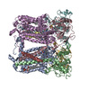
|
|---|---|
| 1 |
|
-Components
-Protein , 4 types, 8 molecules AEBFCGDH
| #1: Protein | Mass: 50087.422 Da / Num. of mol.: 2 / Source method: isolated from a natural source / Source: (natural) Cereibacter sphaeroides (bacteria) / References: UniProt: Q02761#2: Protein | Mass: 29589.158 Da / Num. of mol.: 2 Source method: isolated from a genetically manipulated source Details: Cyt c1 with c-terminal hexa his tag / Source: (gene. exp.) Cereibacter sphaeroides (bacteria) / Gene: petC, fbcC / Production host: #3: Protein | Mass: 19928.375 Da / Num. of mol.: 2 / Source method: isolated from a natural source / Source: (natural) Cereibacter sphaeroides (bacteria) / References: UniProt: Q02762, quinol-cytochrome-c reductase#4: Protein | Mass: 14415.301 Da / Num. of mol.: 2 / Source method: isolated from a natural source Details: This is the natural subunit IV. Only one helix could be determined. Subunit IV has turned out to be rather elusive in numerous crystal structures - it was never determined i.e. got lost ...Details: This is the natural subunit IV. Only one helix could be determined. Subunit IV has turned out to be rather elusive in numerous crystal structures - it was never determined i.e. got lost during crystallization. Here in our CryoEM structure we see it for the first time - even though only one helix Source: (natural) Cereibacter sphaeroides (bacteria) / References: UniProt: P16536 |
|---|
-Non-polymers , 5 types, 12 molecules 
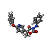


| #5: Chemical | ChemComp-HEM /  Mass: 616.487 Da / Num. of mol.: 4 / Source method: obtained synthetically / Formula: C34H32FeN4O4 Mass: 616.487 Da / Num. of mol.: 4 / Source method: obtained synthetically / Formula: C34H32FeN4O4#6: Chemical |  Mass: 375.377 Da / Num. of mol.: 2 / Source method: obtained synthetically / Formula: C21H17N3O4 / Feature type: SUBJECT OF INVESTIGATION Mass: 375.377 Da / Num. of mol.: 2 / Source method: obtained synthetically / Formula: C21H17N3O4 / Feature type: SUBJECT OF INVESTIGATION#7: Chemical |  Mass: 661.890 Da / Num. of mol.: 2 / Source method: obtained synthetically / Formula: C35H68NO8P / Comment: phospholipid*YM Mass: 661.890 Da / Num. of mol.: 2 / Source method: obtained synthetically / Formula: C35H68NO8P / Comment: phospholipid*YM#8: Chemical |  Mass: 618.503 Da / Num. of mol.: 2 / Source method: obtained synthetically / Formula: C34H34FeN4O4 Mass: 618.503 Da / Num. of mol.: 2 / Source method: obtained synthetically / Formula: C34H34FeN4O4#9: Chemical |  Mass: 175.820 Da / Num. of mol.: 2 / Source method: obtained synthetically / Formula: Fe2S2 Mass: 175.820 Da / Num. of mol.: 2 / Source method: obtained synthetically / Formula: Fe2S2 |
|---|
-Details
| Has ligand of interest | Y |
|---|---|
| Has protein modification | Y |
-Experimental details
-Experiment
| Experiment | Method: ELECTRON MICROSCOPY |
|---|---|
| EM experiment | Aggregation state: PARTICLE / 3D reconstruction method: single particle reconstruction |
- Sample preparation
| Component | Name: Cytochrome bc1 complex of rhodobacter sphaeroides inhibited by pyramoxadone Type: COMPLEX / Entity ID: #1-#4 / Source: NATURAL | |||||||||||||||||||||||||
|---|---|---|---|---|---|---|---|---|---|---|---|---|---|---|---|---|---|---|---|---|---|---|---|---|---|---|
| Molecular weight | Value: 250 kDa/nm / Experimental value: NO | |||||||||||||||||||||||||
| Source (natural) | Organism: Cereibacter sphaeroides (bacteria) | |||||||||||||||||||||||||
| Source (recombinant) | Organism: | |||||||||||||||||||||||||
| Buffer solution | pH: 7.5 | |||||||||||||||||||||||||
| Buffer component |
| |||||||||||||||||||||||||
| Specimen | Conc.: 8 mg/ml / Embedding applied: NO / Shadowing applied: NO / Staining applied: NO / Vitrification applied: YES Details: Cytochrome bc1 complex from Rhodobacter sphaeroides with inhibitor pyramoxadone (PQ4), frozen stock 84 mg/ml. dilute to 8 mg/ml in buffer 50mM TrisHCl, pH7.5/0.16% SMC/0.01% GDN/1mM EDTA | |||||||||||||||||||||||||
| Specimen support | Grid material: COPPER / Grid mesh size: 400 divisions/in. / Grid type: Quantifoil R1.2/1.3 | |||||||||||||||||||||||||
| Vitrification | Instrument: FEI VITROBOT MARK IV / Cryogen name: ETHANE / Humidity: 95 % / Chamber temperature: 277 K Details: 3 ul of sample blot for 3 sec. blot force 20. glow discharge for 60 sec with easy glow |
- Electron microscopy imaging
| Experimental equipment |  Model: Titan Krios / Image courtesy: FEI Company |
|---|---|
| Microscopy | Model: FEI TITAN KRIOS |
| Electron gun | Electron source:  FIELD EMISSION GUN / Accelerating voltage: 300 kV / Illumination mode: FLOOD BEAM FIELD EMISSION GUN / Accelerating voltage: 300 kV / Illumination mode: FLOOD BEAM |
| Electron lens | Mode: BRIGHT FIELD / Nominal magnification: 105000 X / Calibrated magnification: 60241 X / Nominal defocus max: 2500 nm / Nominal defocus min: 1000 nm / Calibrated defocus min: 836 nm / Calibrated defocus max: 3434 nm / Cs: 2.7 mm / C2 aperture diameter: 100 µm / Alignment procedure: COMA FREE |
| Specimen holder | Cryogen: NITROGEN / Specimen holder model: FEI TITAN KRIOS AUTOGRID HOLDER / Temperature (max): 80 K / Temperature (min): 80 K |
| Image recording | Average exposure time: 2.5 sec. / Electron dose: 54.5 e/Å2 / Film or detector model: GATAN K3 BIOQUANTUM (6k x 4k) / Num. of grids imaged: 2 / Num. of real images: 20306 / Details: 0.05s per frame for total 50 frames. |
| EM imaging optics | Energyfilter name: GIF Bioquantum / Energyfilter slit width: 20 eV |
| Image scans | Sampling size: 5 µm / Width: 4092 / Height: 5760 |
- Processing
| Software |
| |||||||||||||||||||||||||||||||||||||||||||||
|---|---|---|---|---|---|---|---|---|---|---|---|---|---|---|---|---|---|---|---|---|---|---|---|---|---|---|---|---|---|---|---|---|---|---|---|---|---|---|---|---|---|---|---|---|---|---|
| EM software |
| |||||||||||||||||||||||||||||||||||||||||||||
| CTF correction | Type: PHASE FLIPPING AND AMPLITUDE CORRECTION | |||||||||||||||||||||||||||||||||||||||||||||
| Particle selection | Num. of particles selected: 5498641 | |||||||||||||||||||||||||||||||||||||||||||||
| Symmetry | Point symmetry: C2 (2 fold cyclic) | |||||||||||||||||||||||||||||||||||||||||||||
| 3D reconstruction | Resolution: 2.91 Å / Resolution method: FSC 0.143 CUT-OFF / Num. of particles: 725256 / Num. of class averages: 2 / Symmetry type: POINT | |||||||||||||||||||||||||||||||||||||||||||||
| Refinement | Cross valid method: NONE Stereochemistry target values: GeoStd + Monomer Library + CDL v1.2 | |||||||||||||||||||||||||||||||||||||||||||||
| Displacement parameters | Biso mean: 56.36 Å2 | |||||||||||||||||||||||||||||||||||||||||||||
| Refine LS restraints |
|
