 Movie
Movie Controller

[English] 日本語
 Yorodumi
- PDB-7tjg: Bacteriophage Q beta capsid protein, A38K/A40C/D102C in T1 symmetry -
+ Open data

- Basic information
| Entry | Database: PDB / ID: 7tjg | ||||||
|---|---|---|---|---|---|---|---|
| Title | Bacteriophage Q beta capsid protein, A38K/A40C/D102C in T1 symmetry | ||||||
 Components Components | Minor capsid protein A1 | ||||||
 Keywords Keywords | VIRUS LIKE PARTICLE / bacteriophage Q beta capsid / T1 symmetry | ||||||
| Function / homology |  Function and homology information Function and homology information | ||||||
| Biological species |  Bacteriophage sp. (virus) Bacteriophage sp. (virus) | ||||||
| Method |  X-RAY DIFFRACTION / SYNCHROTRON / MOLECULAR REPLACEMENT / Resolution: 3.903 Å X-RAY DIFFRACTION / SYNCHROTRON / MOLECULAR REPLACEMENT / Resolution: 3.903 Å | ||||||
 Authors Authors | Jin, X. | ||||||
| Funding support |  United States, 1items United States, 1items
| ||||||
 Citation Citation | Journal: To Be Published Title: Alternative Assembly of Q beta Virus-like Particles Authors: Shaw, V. / Sungsuwan, S. / McFall-Boegeman, H. / Huang, X. / Jin, X. #1: Journal: ACS Chemical Biology / Year: 2022Title: Structure Guided Design of Bacteriophage Q beta Mutants as Next Generation Carriers for Conjugate Vaccines Authors: Sungsuwan, S. / Wu, X. / Shaw, V. / McFall-Boegeman, H. / Rashidijahanabad, Z. / Tan, Z. / Lang, S. / Tahmasebi Nick, S. / Lin, P. / Yin, Z. / Ramadan, S. / Jin, X. / Huang, X. | ||||||
| History |
|
- Structure visualization
| Structure viewer | Molecule: MolmilJmol/JSmol |
|---|
- Downloads & links
-Download
| PDBx/mmCIF format | 7tjg.cif.gz | 132.8 KB | Display | PDBx/mmCIF format |
|---|---|---|---|---|
| PDB format | pdb7tjg.ent.gz | 105.9 KB | Display | PDB format |
| PDBx/mmJSON format | 7tjg.json.gz | Tree view | PDBx/mmJSON format | |
| Others |  Other downloads Other downloads |
-Validation report
| Arichive directory | https://data.pdbj.org/pub/pdb/validation_reports/tj/7tjgftp://data.pdbj.org/pub/pdb/validation_reports/tj/7tjg | HTTPS FTP |
|---|
-Related structure data
| Related structure data |  7tjdC 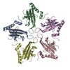 7tjeC 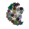 7tjmC  1qbeS C: citing same article ( S: Starting model for refinement |
|---|---|
| Similar structure data |
-Links
 PDBj
PDBj- Assembly
| Deposited unit | 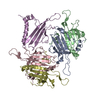
| ||||||||
|---|---|---|---|---|---|---|---|---|---|
| 1 | x 12
| ||||||||
| Unit cell |
|
-Components
| #1: Protein | Mass: 14215.099 Da / Num. of mol.: 5 / Mutation: A38K, A40C, D102C Source method: isolated from a genetically manipulated source Source: (gene. exp.) Bacteriophage sp. (virus) / Production host:  Has protein modification | Y | |
|---|
-Experimental details
-Experiment
| Experiment | Method: X-RAY DIFFRACTION / Number of used crystals: 1 |
|---|
- Sample preparation
| Crystal | Density Matthews: 4.13 Å3/Da / Density % sol: 70.21 % |
|---|---|
| Crystal grow | Temperature: 298 K / Method: vapor diffusion / pH: 7.5 / Details: PEG 3350, L-proline, HEPES / PH range: 7.0-7.5 |
-Data collection
| Diffraction | Mean temperature: 100 K / Serial crystal experiment: N |
|---|---|
| Diffraction source | Source: SYNCHROTRON / Site: APS / Beamline: 21-ID-D / Wavelength: 0.979 Å |
| Detector | Type: DECTRIS EIGER X 16M / Detector: PIXEL / Date: Dec 13, 2017 |
| Radiation | Protocol: SINGLE WAVELENGTH / Monochromatic (M) / Laue (L): M / Scattering type: x-ray |
| Radiation wavelength | Wavelength: 0.979 Å / Relative weight: 1 |
| Reflection | Resolution: 3.9→30.3 Å / Num. obs: 10727 / % possible obs: 98.8 % / Redundancy: 30.6 % / CC1/2: 0.503 / Net I/σ(I): 10.3 |
| Reflection shell | Resolution: 3.9→3.97 Å / Num. unique obs: 1074 / CC1/2: 0.503 |
- Processing
| Software |
| ||||||||||||||||||||||||||||||
|---|---|---|---|---|---|---|---|---|---|---|---|---|---|---|---|---|---|---|---|---|---|---|---|---|---|---|---|---|---|---|---|
| Refinement | Method to determine structure: MOLECULAR REPLACEMENT Starting model: 1qbe Resolution: 3.903→30.3 Å / SU ML: 0.53 / Cross valid method: THROUGHOUT / σ(F): 1.39 / Phase error: 30.61 / Stereochemistry target values: ML
| ||||||||||||||||||||||||||||||
| Solvent computation | Shrinkage radii: 0.9 Å / VDW probe radii: 1.11 Å / Solvent model: FLAT BULK SOLVENT MODEL | ||||||||||||||||||||||||||||||
| Displacement parameters | Biso max: 195.36 Å2 / Biso mean: 120.7138 Å2 / Biso min: 80.37 Å2 | ||||||||||||||||||||||||||||||
| Refinement step | Cycle: final / Resolution: 3.903→30.3 Å
| ||||||||||||||||||||||||||||||
| Refine LS restraints |
| ||||||||||||||||||||||||||||||
| LS refinement shell | Refine-ID: X-RAY DIFFRACTION / Rfactor Rfree error: 0
|
