+ データを開く
- 基本情報
基本情報
| 登録情報 | データベース: PDB / ID: 7thr | ||||||
|---|---|---|---|---|---|---|---|
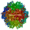
| タイトル | Cryo-electron microscopy of Adeno-associated virus serotype 4 at 2.2 A | ||||||
 要素 要素 | Capsid | ||||||
 キーワード キーワード | VIRUS LIKE PARTICLE / AAV4 / adeno-associated virus / serotype 4 | ||||||
| 機能・相同性 | Phospholipase A2-like domain / Phospholipase A2-like domain / Parvovirus coat protein VP2 / Parvovirus coat protein VP1/VP2 / Parvovirus coat protein VP1/VP2 / Capsid/spike protein, ssDNA virus / T=1 icosahedral viral capsid / structural molecule activity / Capsid 機能・相同性情報 機能・相同性情報 | ||||||
| 生物種 |  Adeno-associated virus - 4 (アデノ随伴ウイルス) Adeno-associated virus - 4 (アデノ随伴ウイルス) | ||||||
| 手法 | 電子顕微鏡法 / 単粒子再構成法 / クライオ電子顕微鏡法 / 解像度: 2.21 Å | ||||||
 データ登録者 データ登録者 | Zane, G.M. / Silveria, M.A. / Meyer, N.L. / White, T.A. / Chapman, M.S. | ||||||
| 資金援助 |  米国, 1件 米国, 1件
| ||||||
 引用 引用 |  ジャーナル: Acta Crystallogr D Struct Biol / 年: 2023 ジャーナル: Acta Crystallogr D Struct Biol / 年: 2023タイトル: Cryo-EM structure of adeno-associated virus 4 at 2.2 Å resolution. 著者: Grant Zane / Mark Silveria / Nancy Meyer / Tommi White / Rui Duan / Xiaoqin Zou / Michael Chapman /  要旨: Adeno-associated virus (AAV) is the vector of choice for several approved gene-therapy treatments and is the basis for many ongoing clinical trials. Various strains of AAV exist (referred to as ...Adeno-associated virus (AAV) is the vector of choice for several approved gene-therapy treatments and is the basis for many ongoing clinical trials. Various strains of AAV exist (referred to as serotypes), each with their own transfection characteristics. Here, a high-resolution cryo-electron microscopy structure (2.2 Å) of AAV serotype 4 (AAV4) is presented. The receptor responsible for transduction of the AAV4 clade of AAV viruses (including AAV11, AAV12 and AAVrh32.33) is unknown. Other AAVs interact with the same cell receptor, adeno-associated virus receptor (AAVR), in one of two different ways. AAV5-like viruses interact exclusively with the polycystic kidney disease-like 1 (PKD1) domain of AAVR, while most other AAVs interact primarily with the PKD2 domain. A comparison of the present AAV4 structure with prior corresponding structures of AAV5, AAV2 and AAV1 in complex with AAVR provides a foundation for understanding why the AAV4-like clade is unable to interact with either PKD1 or PKD2 of AAVR. The conformation of the AAV4 capsid in variable regions I, III, IV and V on the viral surface appears to be sufficiently different from AAV2 to ablate binding with PKD2. Differences between AAV4 and AAV5 in variable region VII appear to be sufficient to exclude binding with PKD1. | ||||||
| 履歴 |
|
- 構造の表示
構造の表示
| 構造ビューア | 分子:  Molmil Molmil Jmol/JSmol Jmol/JSmol |
|---|
- ダウンロードとリンク
- ダウンロード
| PDBx/mmCIF形式 |  7thr.cif.gz 7thr.cif.gz | 125.4 KB | 表示 |  PDBx/mmCIF形式 PDBx/mmCIF形式 |
|---|---|---|---|---|
| PDB形式 |  pdb7thr.ent.gz pdb7thr.ent.gz | 表示 |  PDB形式 PDB形式 | |
| PDBx/mmJSON形式 |  7thr.json.gz 7thr.json.gz | ツリー表示 |  PDBx/mmJSON形式 PDBx/mmJSON形式 | |
| その他 |  その他のダウンロード その他のダウンロード |
-検証レポート
| 文書・要旨 |  7thr_validation.pdf.gz 7thr_validation.pdf.gz | 1.3 MB | 表示 |  wwPDB検証レポート wwPDB検証レポート |
|---|---|---|---|---|
| 文書・詳細版 |  7thr_full_validation.pdf.gz 7thr_full_validation.pdf.gz | 1.3 MB | 表示 | |
| XML形式データ |  7thr_validation.xml.gz 7thr_validation.xml.gz | 41.9 KB | 表示 | |
| CIF形式データ |  7thr_validation.cif.gz 7thr_validation.cif.gz | 60 KB | 表示 | |
| アーカイブディレクトリ |  https://data.pdbj.org/pub/pdb/validation_reports/th/7thr https://data.pdbj.org/pub/pdb/validation_reports/th/7thr ftp://data.pdbj.org/pub/pdb/validation_reports/th/7thr ftp://data.pdbj.org/pub/pdb/validation_reports/th/7thr | HTTPS FTP |
-関連構造データ
| 関連構造データ |  25903MC M: このデータのモデリングに利用したマップデータ C: 同じ文献を引用 ( |
|---|---|
| 類似構造データ | 類似検索 - 機能・相同性  F&H 検索 F&H 検索 |
- リンク
リンク
- 集合体
集合体
| 登録構造単位 | 
|
|---|---|
| 1 | x 60
|
| 2 |
|
| 3 | x 5
|
| 4 | x 6
|
| 5 | 
|
| 対称性 | 点対称性: (シェーンフリース記号: I (正20面体型対称)) |
- 要素
要素
| #1: タンパク質 | 分子量: 80688.023 Da / 分子数: 1 / 変異: M1T / 由来タイプ: 組換発現 / 詳細: VP1 sequence of AAV4 由来: (組換発現)  Adeno-associated virus - 4 (アデノ随伴ウイルス) Adeno-associated virus - 4 (アデノ随伴ウイルス)プラスミド: pFastBac-LIC(4A) / 詳細 (発現宿主): AmpR / 細胞株 (発現宿主): Sf9 発現宿主:  参照: UniProt: O41855 | ||||
|---|---|---|---|---|---|
| #2: 化合物 | | #3: 水 | ChemComp-HOH / | 研究の焦点であるリガンドがあるか | N | |
-実験情報
-実験
| 実験 | 手法: 電子顕微鏡法 |
|---|---|
| EM実験 | 試料の集合状態: PARTICLE / 3次元再構成法: 単粒子再構成法 |
- 試料調製
試料調製
| 構成要素 | 名称: Adeno-associated virus / タイプ: VIRUS / Entity ID: #1 / 由来: RECOMBINANT | |||||||||||||||
|---|---|---|---|---|---|---|---|---|---|---|---|---|---|---|---|---|
| 分子量 | 値: 3.746 MDa / 実験値: NO | |||||||||||||||
| 由来(天然) | 生物種:   Adeno-associated virus (アデノ随伴ウイルス) Adeno-associated virus (アデノ随伴ウイルス)株: Serotype 4 | |||||||||||||||
| 由来(組換発現) | 生物種:  株: Sf9 / プラスミド: pFastBac-LIC(4A) | |||||||||||||||
| ウイルスについての詳細 | 中空か: YES / エンベロープを持つか: NO / 単離: SEROTYPE / タイプ: VIRUS-LIKE PARTICLE | |||||||||||||||
| 天然宿主 | 生物種: Homo sapiens | |||||||||||||||
| ウイルス殻 | 名称: Capsid / 直径: 250 nm / 三角数 (T数): 1 | |||||||||||||||
| 緩衝液 | pH: 7.4 | |||||||||||||||
| 緩衝液成分 |
| |||||||||||||||
| 試料 | 濃度: 0.33 mg/ml / 包埋: NO / シャドウイング: NO / 染色: NO / 凍結: YES 詳細: The sample was isolated by multiple rounds of cesium chloride density centrifugation and dialyzed into a HEPES-buffered, sodium chloride solution (see Meyer et al., 2019 Bioprotocols paper). | |||||||||||||||
| 試料支持 | グリッドの材料: COPPER / グリッドのサイズ: 400 divisions/in. グリッドのタイプ: PELCO Ultrathin Carbon with Lacey Carbon | |||||||||||||||
| 急速凍結 | 装置: FEI VITROBOT MARK IV / 凍結剤: ETHANE / 湿度: 100 % / 凍結前の試料温度: 298 K / 詳細: blot force: 4 time: 2 s |
- 電子顕微鏡撮影
| 実験機器 |  モデル: Titan Krios / 画像提供: FEI Company |
|---|---|
| 顕微鏡 | モデル: FEI TITAN KRIOS |
| 電子銃 | 電子線源:  FIELD EMISSION GUN / 加速電圧: 300 kV / 照射モード: FLOOD BEAM FIELD EMISSION GUN / 加速電圧: 300 kV / 照射モード: FLOOD BEAM |
| 電子レンズ | モード: BRIGHT FIELD / 倍率(公称値): 16500 X / 最大 デフォーカス(公称値): 1800 nm / 最小 デフォーカス(公称値): 600 nm / C2レンズ絞り径: 50 µm |
| 撮影 | 平均露光時間: 1.4 sec. / 電子線照射量: 48 e/Å2 フィルム・検出器のモデル: GATAN K3 BIOQUANTUM (6k x 4k) 撮影したグリッド数: 1 / 実像数: 4809 |
| 電子光学装置 | エネルギーフィルター名称: GIF Bioquantum / エネルギーフィルタースリット幅: 20 eV |
- 解析
解析
| EMソフトウェア |
| |||||||||||||||||||||||||||||||||||||||||||||
|---|---|---|---|---|---|---|---|---|---|---|---|---|---|---|---|---|---|---|---|---|---|---|---|---|---|---|---|---|---|---|---|---|---|---|---|---|---|---|---|---|---|---|---|---|---|---|
| CTF補正 | タイプ: PHASE FLIPPING ONLY | |||||||||||||||||||||||||||||||||||||||||||||
| 粒子像の選択 | 選択した粒子像数: 75226 | |||||||||||||||||||||||||||||||||||||||||||||
| 対称性 | 点対称性: I (正20面体型対称) | |||||||||||||||||||||||||||||||||||||||||||||
| 3次元再構成 | 解像度: 2.21 Å / 解像度の算出法: FSC 0.143 CUT-OFF / 粒子像の数: 75226 / アルゴリズム: FOURIER SPACE / クラス平均像の数: 1 / 対称性のタイプ: POINT | |||||||||||||||||||||||||||||||||||||||||||||
| 原子モデル構築 | 詳細: Link to CNS_RSRef software: https://chapman.missouri.edu/software/ | |||||||||||||||||||||||||||||||||||||||||||||
| 原子モデル構築 | PDB-ID: 2G8G PDB chain-ID: A / Accession code: 2G8G / Pdb chain residue range: 211-734 / Source name: PDB / タイプ: experimental model |
 ムービー
ムービー コントローラー


 PDBj
PDBj


