 Movie
Movie Controller


+ Open data

- Basic information
| Entry | Database: PDB / ID: 7th5 | ||||||
|---|---|---|---|---|---|---|---|
| Title | Thermus thermophilus methylenetetrahydrofolate reductase | ||||||
 Components Components | Methylenetetrahydrofolate reductase | ||||||
 Keywords Keywords | OXIDOREDUCTASE / TIM barrel / flavin | ||||||
| Function / homology |  Function and homology information Function and homology informationmethylenetetrahydrofolate reductase (NADH) / methylenetetrahydrofolate reductase (NADH) activity / : / tetrahydrofolate interconversion / FAD binding / cytosol Similarity search - Function | ||||||
| Biological species |   Thermus thermophilus HB8 (bacteria) Thermus thermophilus HB8 (bacteria) | ||||||
| Method |  X-RAY DIFFRACTION / SYNCHROTRON / MOLECULAR REPLACEMENT / Resolution: 2.09 Å X-RAY DIFFRACTION / SYNCHROTRON / MOLECULAR REPLACEMENT / Resolution: 2.09 Å | ||||||
 Authors Authors | Yamada, K. / Koutmos, M. | ||||||
| Funding support |  United States, 1items United States, 1items
| ||||||
 Citation Citation | Journal: J.Biol.Chem. / Year: 2022 Title: 5-Formyltetrahydrofolate promotes conformational remodeling in a methylenetetrahydrofolate reductase active site and inhibits its activity. Authors: Yamada, K. / Mendoza, J. / Koutmos, M. | ||||||
| History |
|
- Structure visualization
| Structure viewer | Molecule: MolmilJmol/JSmol |
|---|
- Downloads & links
-Download
| PDBx/mmCIF format | 7th5.cif.gz | 233.7 KB | Display | PDBx/mmCIF format |
|---|---|---|---|---|
| PDB format | pdb7th5.ent.gz | 188.9 KB | Display | PDB format |
| PDBx/mmJSON format | 7th5.json.gz | Tree view | PDBx/mmJSON format | |
| Others |  Other downloads Other downloads |
-Validation report
| Arichive directory | https://data.pdbj.org/pub/pdb/validation_reports/th/7th5ftp://data.pdbj.org/pub/pdb/validation_reports/th/7th5 | HTTPS FTP |
|---|
-Related structure data
| Related structure data | 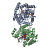 7th4C  8eacC 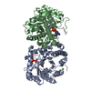 3apyS S: Starting model for refinement C: citing same article ( |
|---|---|
| Similar structure data |
-Links
 PDBj
PDBj- Assembly
| Deposited unit | 
| ||||||||
|---|---|---|---|---|---|---|---|---|---|
| 1 |
| ||||||||
| Unit cell |
|
-Components
| #1: Protein | Mass: 33050.887 Da / Num. of mol.: 2 Source method: isolated from a genetically manipulated source Source: (gene. exp.) Thermus thermophilus HB8 (bacteria) / Strain: ATCC 27634 / DSM 579 / HB8 / Gene: TTHA0327 / Production host: References: UniProt: Q5SLG6, methylenetetrahydrofolate reductase [NAD(P)H] #2: Chemical |   Mass: 785.550 Da / Num. of mol.: 2 / Source method: obtained synthetically / Formula: C27H33N9O15P2 / Feature type: SUBJECT OF INVESTIGATION / Comment: FAD*YM Mass: 785.550 Da / Num. of mol.: 2 / Source method: obtained synthetically / Formula: C27H33N9O15P2 / Feature type: SUBJECT OF INVESTIGATION / Comment: FAD*YM#3: Chemical |   Mass: 22.990 Da / Num. of mol.: 2 / Source method: obtained synthetically / Formula: Na Mass: 22.990 Da / Num. of mol.: 2 / Source method: obtained synthetically / Formula: Na#4: Water | ChemComp-HOH / |  Mass: 18.015 Da / Num. of mol.: 156 / Source method: isolated from a natural source / Formula: H2O Mass: 18.015 Da / Num. of mol.: 156 / Source method: isolated from a natural source / Formula: H2OHas ligand of interest | Y | |
|---|
-Experimental details
-Experiment
| Experiment | Method: X-RAY DIFFRACTION / Number of used crystals: 1 |
|---|
- Sample preparation
| Crystal | Density Matthews: 2.72 Å3/Da / Density % sol: 54.79 % |
|---|---|
| Crystal grow | Temperature: 297 K / Method: vapor diffusion, sitting drop / pH: 8.5 / Details: 0.1M Tris-HCl pH 8.5, 3M NaCl |
-Data collection
| Diffraction | Mean temperature: 100 K / Serial crystal experiment: N |
|---|---|
| Diffraction source | Source: SYNCHROTRON / Site: APS / Beamline: 23-ID-D / Wavelength: 1.0332 Å |
| Detector | Type: DECTRIS PILATUS3 6M / Detector: PIXEL / Date: Jun 28, 2018 |
| Radiation | Protocol: SINGLE WAVELENGTH / Monochromatic (M) / Laue (L): M / Scattering type: x-ray |
| Radiation wavelength | Wavelength: 1.0332 Å / Relative weight: 1 |
| Reflection | Resolution: 2.09→47.07 Å / Num. obs: 42742 / % possible obs: 98.7 % / Redundancy: 3.9 % / CC1/2: 0.993 / Rmerge(I) obs: 0.118 / Rpim(I) all: 0.069 / Rrim(I) all: 0.138 / Net I/σ(I): 7.6 |
| Reflection shell | Resolution: 2.09→2.15 Å / Redundancy: 3.5 % / Rmerge(I) obs: 0.621 / Num. unique obs: 2924 / CC1/2: 0.674 / Rpim(I) all: 0.316 / Rrim(I) all: 0.621 / % possible all: 88.4 |
- Processing
| Software |
| ||||||||||||||||||||||||||||||||||||||||||||||||||||||||||||
|---|---|---|---|---|---|---|---|---|---|---|---|---|---|---|---|---|---|---|---|---|---|---|---|---|---|---|---|---|---|---|---|---|---|---|---|---|---|---|---|---|---|---|---|---|---|---|---|---|---|---|---|---|---|---|---|---|---|---|---|---|---|
| Refinement | Method to determine structure: MOLECULAR REPLACEMENT Starting model: 3APY Resolution: 2.09→47.07 Å / Cor.coef. Fo:Fc: 0.955 / Cor.coef. Fo:Fc free: 0.934 / SU B: 5.074 / SU ML: 0.128 / Cross valid method: THROUGHOUT / σ(F): 0 / ESU R: 0.185 / ESU R Free: 0.165 / Stereochemistry target values: MAXIMUM LIKELIHOOD Details: HYDROGENS HAVE BEEN USED IF PRESENT IN THE INPUT U VALUES : REFINED INDIVIDUALLY
| ||||||||||||||||||||||||||||||||||||||||||||||||||||||||||||
| Solvent computation | Ion probe radii: 0.8 Å / Shrinkage radii: 0.8 Å / VDW probe radii: 1.2 Å / Solvent model: BABINET MODEL WITH MASK | ||||||||||||||||||||||||||||||||||||||||||||||||||||||||||||
| Displacement parameters | Biso max: 218.89 Å2 / Biso mean: 34.665 Å2 / Biso min: 10.84 Å2
| ||||||||||||||||||||||||||||||||||||||||||||||||||||||||||||
| Refinement step | Cycle: final / Resolution: 2.09→47.07 Å
| ||||||||||||||||||||||||||||||||||||||||||||||||||||||||||||
| Refine LS restraints |
| ||||||||||||||||||||||||||||||||||||||||||||||||||||||||||||
| LS refinement shell | Resolution: 2.094→2.149 Å / Rfactor Rfree error: 0 / Total num. of bins used: 20
|
