 Movie
Movie Controller

[English] 日本語
 Yorodumi
- PDB-7tef: Cytochrome P450 14 alpha-sterol demethylase CYP51 from Mycobacter... -
+ Open data

- Basic information
| Entry | Database: PDB / ID: 7tef | ||||||
|---|---|---|---|---|---|---|---|
| Title | Cytochrome P450 14 alpha-sterol demethylase CYP51 from Mycobacterium marinum | ||||||
 Components Components | Cytochrome P450 51B1 Cyp51B1 | ||||||
 Keywords Keywords | OXIDOREDUCTASE / 14 alpha sterol demethylase Cytochrome P450 Substrate free | ||||||
| Function / homology |  Function and homology information Function and homology informationoxidoreductase activity, acting on paired donors, with incorporation or reduction of molecular oxygen / monooxygenase activity / iron ion binding / heme binding Similarity search - Function | ||||||
| Biological species |  Mycobacterium marinum (bacteria) Mycobacterium marinum (bacteria) | ||||||
| Method |  X-RAY DIFFRACTION / SYNCHROTRON / MOLECULAR REPLACEMENT / molecular replacement / Resolution: 1.98 Å X-RAY DIFFRACTION / SYNCHROTRON / MOLECULAR REPLACEMENT / molecular replacement / Resolution: 1.98 Å | ||||||
 Authors Authors | Mohamed, H.A. / Bruning, J.B. / Bell, S.G. | ||||||
| Funding support |  Australia, 1items Australia, 1items
| ||||||
 Citation Citation | Journal: J.Steroid Biochem.Mol.Biol. / Year: 2022 Title: A comparison of the bacterial CYP51 cytochrome P450 enzymes from Mycobacterium marinum and Mycobacterium tuberculosis. Authors: Mohamed, H. / Child, S.A. / Bruning, J.B. / Bell, S.G. | ||||||
| History |
|
- Structure visualization
| Structure viewer | Molecule: MolmilJmol/JSmol |
|---|
- Downloads & links
-Download
| PDBx/mmCIF format | 7tef.cif.gz | 119 KB | Display | PDBx/mmCIF format |
|---|---|---|---|---|
| PDB format | pdb7tef.ent.gz | 87.2 KB | Display | PDB format |
| PDBx/mmJSON format | 7tef.json.gz | Tree view | PDBx/mmJSON format | |
| Others |  Other downloads Other downloads |
-Validation report
| Summary document | 7tef_validation.pdf.gz | 2.8 MB | Display | wwPDB validaton report |
|---|---|---|---|---|
| Full document | 7tef_full_validation.pdf.gz | 2.8 MB | Display | |
| Data in XML | 7tef_validation.xml.gz | 23.1 KB | Display | |
| Data in CIF | 7tef_validation.cif.gz | 35.2 KB | Display | |
| Arichive directory | https://data.pdbj.org/pub/pdb/validation_reports/te/7tefftp://data.pdbj.org/pub/pdb/validation_reports/te/7tef | HTTPS FTP |
-Related structure data
| Related structure data |  1ea1S S: Starting model for refinement |
|---|---|
| Similar structure data |
-Links
 PDBj
PDBj
- Assembly
| Deposited unit | 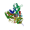
| ||||||||
|---|---|---|---|---|---|---|---|---|---|
| 1 |
| ||||||||
| Unit cell |
|
-Components
| #1: Protein | Mass: 52441.270 Da / Num. of mol.: 1 Source method: isolated from a genetically manipulated source Source: (gene. exp.) Mycobacterium marinum (bacteria) / Strain: ATCC BAA-535 / M / Gene: cyp51B1, MMAR_4932 / Production host: | ||||
|---|---|---|---|---|---|
| #2: Chemical | ChemComp-HEM /   Mass: 616.487 Da / Num. of mol.: 1 / Source method: obtained synthetically / Formula: C34H32FeN4O4 / Feature type: SUBJECT OF INVESTIGATION Mass: 616.487 Da / Num. of mol.: 1 / Source method: obtained synthetically / Formula: C34H32FeN4O4 / Feature type: SUBJECT OF INVESTIGATION | ||||
| #3: Chemical | ChemComp-BTB /   Mass: 209.240 Da / Num. of mol.: 1 / Source method: obtained synthetically / Formula: C8H19NO5 / Feature type: SUBJECT OF INVESTIGATION / Comment: pH buffer*YM Mass: 209.240 Da / Num. of mol.: 1 / Source method: obtained synthetically / Formula: C8H19NO5 / Feature type: SUBJECT OF INVESTIGATION / Comment: pH buffer*YM | ||||
| #4: Chemical | ChemComp-SO4 /   Mass: 96.063 Da / Num. of mol.: 5 / Source method: obtained synthetically / Formula: SO4 / Feature type: SUBJECT OF INVESTIGATION Mass: 96.063 Da / Num. of mol.: 5 / Source method: obtained synthetically / Formula: SO4 / Feature type: SUBJECT OF INVESTIGATION#5: Water | ChemComp-HOH / |  Mass: 18.015 Da / Num. of mol.: 454 / Source method: isolated from a natural source / Formula: H2O Mass: 18.015 Da / Num. of mol.: 454 / Source method: isolated from a natural source / Formula: H2OHas ligand of interest | Y | |
-Experimental details
-Experiment
| Experiment | Method: X-RAY DIFFRACTION / Number of used crystals: 1 |
|---|
- Sample preparation
| Crystal | Density Matthews: 2.24 Å3/Da / Density meas: 1 Mg/m3 / Density % sol: 45.04 % / Description: Needle-shaped crystals |
|---|---|
| Crystal grow | Temperature: 289.15 K / Method: vapor diffusion / pH: 6.5 / Details: Bis-Tris, PEG 3350, ammonium sulfate / PH range: 5.5-6.5 |
-Data collection
| Diffraction | Mean temperature: 100 K / Serial crystal experiment: N | ||||||||||||||||||||||||
|---|---|---|---|---|---|---|---|---|---|---|---|---|---|---|---|---|---|---|---|---|---|---|---|---|---|
| Diffraction source | Source: SYNCHROTRON / Site: Australian Synchrotron / Beamline: MX1 / Wavelength: 1 Å | ||||||||||||||||||||||||
| Detector | Type: ADSC QUANTUM 1 / Detector: CCD / Date: Jun 14, 2020 | ||||||||||||||||||||||||
| Radiation | Protocol: SINGLE WAVELENGTH / Monochromatic (M) / Laue (L): M / Scattering type: x-ray | ||||||||||||||||||||||||
| Radiation wavelength | Wavelength: 1 Å / Relative weight: 1 | ||||||||||||||||||||||||
| Reflection | Resolution: 1.98→44.28 Å / Num. obs: 33253 / % possible obs: 100 % / Redundancy: 13.5 % / CC1/2: 0.996 / Rmerge(I) obs: 0.27 / Net I/σ(I): 9.3 / Num. measured all: 447875 / Scaling rejects: 1 | ||||||||||||||||||||||||
| Reflection shell | Diffraction-ID: 1
|
-Phasing
| Phasing | Method: molecular replacement |
|---|
- Processing
| Software |
| ||||||||||||||||||||||||||||||||||||||||||||||||||||||||||||||||||||||||||||||
|---|---|---|---|---|---|---|---|---|---|---|---|---|---|---|---|---|---|---|---|---|---|---|---|---|---|---|---|---|---|---|---|---|---|---|---|---|---|---|---|---|---|---|---|---|---|---|---|---|---|---|---|---|---|---|---|---|---|---|---|---|---|---|---|---|---|---|---|---|---|---|---|---|---|---|---|---|---|---|---|
| Refinement | Method to determine structure: MOLECULAR REPLACEMENT Starting model: 1EA1 Resolution: 1.98→44.28 Å / SU ML: 0.2 / Cross valid method: THROUGHOUT / σ(F): 1.34 / Phase error: 20.23 / Stereochemistry target values: ML
| ||||||||||||||||||||||||||||||||||||||||||||||||||||||||||||||||||||||||||||||
| Solvent computation | Shrinkage radii: 0.9 Å / VDW probe radii: 1.11 Å / Solvent model: FLAT BULK SOLVENT MODEL | ||||||||||||||||||||||||||||||||||||||||||||||||||||||||||||||||||||||||||||||
| Displacement parameters | Biso max: 126.78 Å2 / Biso mean: 26.3023 Å2 / Biso min: 9.24 Å2 | ||||||||||||||||||||||||||||||||||||||||||||||||||||||||||||||||||||||||||||||
| Refinement step | Cycle: final / Resolution: 1.98→44.28 Å
| ||||||||||||||||||||||||||||||||||||||||||||||||||||||||||||||||||||||||||||||
| LS refinement shell | Refine-ID: X-RAY DIFFRACTION / Rfactor Rfree error: 0 / Total num. of bins used: 12 / % reflection obs: 100 %
|
