 Movie
Movie Controller

[English] 日本語
 Yorodumi
- PDB-7ted: Human Ornithine Aminotransferase cocrystallized with its inhibito... -
+ Open data

- Basic information
| Entry | Database: PDB / ID: 7ted | |||||||||
|---|---|---|---|---|---|---|---|---|---|---|
| Title | Human Ornithine Aminotransferase cocrystallized with its inhibitor, (S,E)-3-amino-4-(fluoromethylene)cyclopent-1-ene-1-carboxylate | |||||||||
 Components Components | Ornithine aminotransferase, mitochondrial | |||||||||
 Keywords Keywords | TRANSFERASE/Inhibitor / Human Ornithine Aminotransferase / hOAT / OAT / mechanism-based inhibitor / MBI / irreversible inhibitor / inactivator / TRANSFERASE / TRANSFERASE-Inhibitor complex | |||||||||
| Function / homology |  Function and homology information Function and homology information: / ornithine aminotransferase / L-ornithine transaminase activity / : / L-proline biosynthetic process / Glutamate and glutamine metabolism / visual perception / pyridoxal phosphate binding / mitochondrial matrix / mitochondrion ...: / ornithine aminotransferase / L-ornithine transaminase activity / : / L-proline biosynthetic process / Glutamate and glutamine metabolism / visual perception / pyridoxal phosphate binding / mitochondrial matrix / mitochondrion / nucleoplasm / identical protein binding / cytoplasm Similarity search - Function | |||||||||
| Biological species |  Homo sapiens (human) Homo sapiens (human) | |||||||||
| Method |  X-RAY DIFFRACTION / SYNCHROTRON / MOLECULAR REPLACEMENT / Resolution: 2.63 Å X-RAY DIFFRACTION / SYNCHROTRON / MOLECULAR REPLACEMENT / Resolution: 2.63 Å | |||||||||
 Authors Authors | Butrin, A. / Zhu, W. / Silverman, R. / Liu, D. | |||||||||
| Funding support |  United States, 2items United States, 2items
| |||||||||
 Citation Citation | Journal: J.Am.Chem.Soc. / Year: 2022 Title: Rational Design, Synthesis, and Mechanism of (3 S ,4 R )-3-Amino-4-(difluoromethyl)cyclopent-1-ene-1-carboxylic Acid: Employing a Second-Deprotonation Strategy for Selectivity of Human ...Title: Rational Design, Synthesis, and Mechanism of (3 S ,4 R )-3-Amino-4-(difluoromethyl)cyclopent-1-ene-1-carboxylic Acid: Employing a Second-Deprotonation Strategy for Selectivity of Human Ornithine Aminotransferase over GABA Aminotransferase. Authors: Zhu, W. / Butrin, A. / Melani, R.D. / Doubleday, P.F. / Ferreira, G.M. / Tavares, M.T. / Habeeb Mohammad, T.S. / Beaupre, B.A. / Kelleher, N.L. / Moran, G.R. / Liu, D. / Silverman, R.B. | |||||||||
| History |
|
- Structure visualization
| Structure viewer | Molecule: MolmilJmol/JSmol |
|---|
- Downloads & links
-Download
| PDBx/mmCIF format | 7ted.cif.gz | 694.9 KB | Display | PDBx/mmCIF format |
|---|---|---|---|---|
| PDB format | pdb7ted.ent.gz | 571.7 KB | Display | PDB format |
| PDBx/mmJSON format | 7ted.json.gz | Tree view | PDBx/mmJSON format | |
| Others |  Other downloads Other downloads |
-Validation report
| Arichive directory | https://data.pdbj.org/pub/pdb/validation_reports/te/7tedftp://data.pdbj.org/pub/pdb/validation_reports/te/7ted | HTTPS FTP |
|---|
-Related structure data
| Related structure data |  7tevC  7tfpC 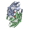 1oatS S: Starting model for refinement C: citing same article ( |
|---|---|
| Similar structure data |
-Links
 PDBj
PDBj- Assembly
| Deposited unit | 
| ||||||||||||
|---|---|---|---|---|---|---|---|---|---|---|---|---|---|
| 1 | 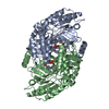
| ||||||||||||
| 2 | 
| ||||||||||||
| 3 | 
| ||||||||||||
| 4 | 
| ||||||||||||
| 5 | 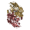
| ||||||||||||
| Unit cell |
| ||||||||||||
| Components on special symmetry positions |
|
-Components
| #1: Protein | Mass: 44860.320 Da / Num. of mol.: 9 Source method: isolated from a genetically manipulated source Source: (gene. exp.) Homo sapiens (human) / Gene: OAT / Production host:  #2: Chemical | ChemComp-I1T / (   Mass: 388.310 Da / Num. of mol.: 8 / Source method: obtained synthetically / Formula: C15H21N2O8P / Feature type: SUBJECT OF INVESTIGATION Mass: 388.310 Da / Num. of mol.: 8 / Source method: obtained synthetically / Formula: C15H21N2O8P / Feature type: SUBJECT OF INVESTIGATION#3: Chemical | ChemComp-PLP / |   Mass: 247.142 Da / Num. of mol.: 1 / Source method: obtained synthetically / Formula: C8H10NO6P / Feature type: SUBJECT OF INVESTIGATION Mass: 247.142 Da / Num. of mol.: 1 / Source method: obtained synthetically / Formula: C8H10NO6P / Feature type: SUBJECT OF INVESTIGATION#4: Water | ChemComp-HOH / |  Mass: 18.015 Da / Num. of mol.: 335 / Source method: isolated from a natural source / Formula: H2O Mass: 18.015 Da / Num. of mol.: 335 / Source method: isolated from a natural source / Formula: H2OHas ligand of interest | Y | |
|---|
-Experimental details
-Experiment
| Experiment | Method: X-RAY DIFFRACTION / Number of used crystals: 1 |
|---|
- Sample preparation
| Crystal | Density Matthews: 2.65 Å3/Da / Density % sol: 53.54 % |
|---|---|
| Crystal grow | Temperature: 293 K / Method: vapor diffusion, hanging drop / pH: 7.8 Details: After purification, OAT was buffer exchanged into the crystallization buffer (50 mM Tricine pH 7.8) supplied supplemented with 1 mM 2-ketoglutarate. The protein was concentrated to 6.5 mg/mL. ...Details: After purification, OAT was buffer exchanged into the crystallization buffer (50 mM Tricine pH 7.8) supplied supplemented with 1 mM 2-ketoglutarate. The protein was concentrated to 6.5 mg/mL. Previously reported crystallization conditions were optimized using the hanging drop vapor diffusion method by varying PEG 6000 (8-12%), NaCl (100-250 mM), and glycerol (0%-10%) with 100 mM Tricine pH 7.8 was being kept constant as the buffer. For each hanging drop, 2 uL of protein solution was mixed with an equal volume of well solution and 0.5 uL of ligand. The crystals with the best morphology and size grew in a final condition containing 12% PEG 6000, 200 mM NaCl, 10% glycerol, and 100 mM Tricine pH 7.8. Crystals were transferred to a cryo-protectant solution (well solution supplemented with 30% glycerol) and flash-frozen in liquid nitrogen |
-Data collection
| Diffraction | Mean temperature: 100 K / Serial crystal experiment: N |
|---|---|
| Diffraction source | Source: SYNCHROTRON / Site: APS / Beamline: 21-ID-D / Wavelength: 1.127 Å |
| Detector | Type: DECTRIS EIGER X 9M / Detector: PIXEL / Date: Dec 13, 2020 |
| Radiation | Protocol: SINGLE WAVELENGTH / Monochromatic (M) / Laue (L): M / Scattering type: x-ray |
| Radiation wavelength | Wavelength: 1.127 Å / Relative weight: 1 |
| Reflection | Resolution: 2.63→43.73 Å / Num. obs: 123624 / % possible obs: 98.9 % / Redundancy: 4.7 % / Biso Wilson estimate: 46.48 Å2 / CC1/2: 0.987 / Net I/σ(I): 8.2 |
| Reflection shell | Resolution: 2.63→2.7 Å / Mean I/σ(I) obs: 1.7 / Num. unique obs: 9062 / CC1/2: 0.294 |
- Processing
| Software |
| |||||||||||||||||||||||||||||||||||||||||||||||||||||||||||||||||||||||||||||||||||||||||||||||||||||||||||||||||||||||||||||||||||||||||||||||||||||||||||||||||||||||||||||||||||||||||||||||||||||||||||||||||||||||||
|---|---|---|---|---|---|---|---|---|---|---|---|---|---|---|---|---|---|---|---|---|---|---|---|---|---|---|---|---|---|---|---|---|---|---|---|---|---|---|---|---|---|---|---|---|---|---|---|---|---|---|---|---|---|---|---|---|---|---|---|---|---|---|---|---|---|---|---|---|---|---|---|---|---|---|---|---|---|---|---|---|---|---|---|---|---|---|---|---|---|---|---|---|---|---|---|---|---|---|---|---|---|---|---|---|---|---|---|---|---|---|---|---|---|---|---|---|---|---|---|---|---|---|---|---|---|---|---|---|---|---|---|---|---|---|---|---|---|---|---|---|---|---|---|---|---|---|---|---|---|---|---|---|---|---|---|---|---|---|---|---|---|---|---|---|---|---|---|---|---|---|---|---|---|---|---|---|---|---|---|---|---|---|---|---|---|---|---|---|---|---|---|---|---|---|---|---|---|---|---|---|---|---|---|---|---|---|---|---|---|---|---|---|---|---|---|---|---|---|
| Refinement | Method to determine structure: MOLECULAR REPLACEMENT Starting model: 1OAT Resolution: 2.63→43.4 Å / SU ML: 0.4167 / Cross valid method: FREE R-VALUE / σ(F): 1.36 / Phase error: 29.5568 Stereochemistry target values: GeoStd + Monomer Library + CDL v1.2
| |||||||||||||||||||||||||||||||||||||||||||||||||||||||||||||||||||||||||||||||||||||||||||||||||||||||||||||||||||||||||||||||||||||||||||||||||||||||||||||||||||||||||||||||||||||||||||||||||||||||||||||||||||||||||
| Solvent computation | Shrinkage radii: 0.9 Å / VDW probe radii: 1.11 Å / Solvent model: FLAT BULK SOLVENT MODEL | |||||||||||||||||||||||||||||||||||||||||||||||||||||||||||||||||||||||||||||||||||||||||||||||||||||||||||||||||||||||||||||||||||||||||||||||||||||||||||||||||||||||||||||||||||||||||||||||||||||||||||||||||||||||||
| Displacement parameters | Biso mean: 18.93 Å2 | |||||||||||||||||||||||||||||||||||||||||||||||||||||||||||||||||||||||||||||||||||||||||||||||||||||||||||||||||||||||||||||||||||||||||||||||||||||||||||||||||||||||||||||||||||||||||||||||||||||||||||||||||||||||||
| Refinement step | Cycle: LAST / Resolution: 2.63→43.4 Å
| |||||||||||||||||||||||||||||||||||||||||||||||||||||||||||||||||||||||||||||||||||||||||||||||||||||||||||||||||||||||||||||||||||||||||||||||||||||||||||||||||||||||||||||||||||||||||||||||||||||||||||||||||||||||||
| Refine LS restraints |
| |||||||||||||||||||||||||||||||||||||||||||||||||||||||||||||||||||||||||||||||||||||||||||||||||||||||||||||||||||||||||||||||||||||||||||||||||||||||||||||||||||||||||||||||||||||||||||||||||||||||||||||||||||||||||
| LS refinement shell |
|
