 Movie
Movie Controller


+ Open data

- Basic information
| Entry | Database: PDB / ID: 7te2 | ||||||
|---|---|---|---|---|---|---|---|
| Title | Crystal Structure of AerR from Rhodobacter capsulatus at 2.25 A. | ||||||
 Components Components | AerR | ||||||
 Keywords Keywords | TRANSCRIPTION / B12 binding / transcription regulator / photosynthesis / gene regulator | ||||||
| Function / homology | B12 binding domain / Cobalamin-binding domain superfamily / B12-binding domain profile. / Cobalamin (vitamin B12)-binding domain / cobalamin binding / metal ion binding / COBALAMIN / HEXANE-1,6-DIOL / B12-binding domain-containing protein Function and homology information Function and homology information | ||||||
| Biological species |  Rhodobacter capsulatus Y262 (bacteria) Rhodobacter capsulatus Y262 (bacteria) | ||||||
| Method |  X-RAY DIFFRACTION / SYNCHROTRON / SAD / Resolution: 2.25 Å X-RAY DIFFRACTION / SYNCHROTRON / SAD / Resolution: 2.25 Å | ||||||
 Authors Authors | Dragnea, V. / Gonzalez-Gutierrez, G. / Bauer, C.E. | ||||||
| Funding support |  United States, 1items United States, 1items
| ||||||
 Citation Citation | Journal: Microorganisms / Year: 2022 Title: Structural Analyses of CrtJ and Its B 12 -Binding Co-Regulators SAerR and LAerR from the Purple Photosynthetic Bacterium Rhodobacter capsulatus. Authors: Dragnea, V. / Gonzalez-Gutierrez, G. / Bauer, C.E. | ||||||
| History |
|
- Structure visualization
| Structure viewer | Molecule: MolmilJmol/JSmol |
|---|
- Downloads & links
-Download
| PDBx/mmCIF format | 7te2.cif.gz | 121.3 KB | Display | PDBx/mmCIF format |
|---|---|---|---|---|
| PDB format | pdb7te2.ent.gz | 77.3 KB | Display | PDB format |
| PDBx/mmJSON format | 7te2.json.gz | Tree view | PDBx/mmJSON format | |
| Others |  Other downloads Other downloads |
-Validation report
| Arichive directory | https://data.pdbj.org/pub/pdb/validation_reports/te/7te2ftp://data.pdbj.org/pub/pdb/validation_reports/te/7te2 | HTTPS FTP |
|---|
-Related structure data
| Similar structure data |
|---|
-Links
 PDBj
PDBj- Assembly
| Deposited unit | 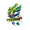
| ||||||||||||
|---|---|---|---|---|---|---|---|---|---|---|---|---|---|
| 1 |
| ||||||||||||
| Unit cell |
| ||||||||||||
| Components on special symmetry positions |
|
-Components
| #1: Protein | Mass: 22754.447 Da / Num. of mol.: 1 Source method: isolated from a genetically manipulated source Source: (gene. exp.) Rhodobacter capsulatus Y262 (bacteria) / Gene: U713_07140 / Production host: | ||||||
|---|---|---|---|---|---|---|---|
| #2: Chemical | ChemComp-B12 /   Mass: 1330.356 Da / Num. of mol.: 1 / Source method: obtained synthetically / Formula: C62H89CoN13O14P / Feature type: SUBJECT OF INVESTIGATION Mass: 1330.356 Da / Num. of mol.: 1 / Source method: obtained synthetically / Formula: C62H89CoN13O14P / Feature type: SUBJECT OF INVESTIGATION | ||||||
| #3: Chemical | ChemComp-HEZ /   Mass: 118.174 Da / Num. of mol.: 1 / Source method: obtained synthetically / Formula: C6H14O2 Mass: 118.174 Da / Num. of mol.: 1 / Source method: obtained synthetically / Formula: C6H14O2 | ||||||
| #4: Chemical |   Mass: 35.453 Da / Num. of mol.: 3 / Source method: obtained synthetically / Formula: Cl Mass: 35.453 Da / Num. of mol.: 3 / Source method: obtained synthetically / Formula: Cl#5: Water | ChemComp-HOH / |  Mass: 18.015 Da / Num. of mol.: 77 / Source method: isolated from a natural source / Formula: H2O Mass: 18.015 Da / Num. of mol.: 77 / Source method: isolated from a natural source / Formula: H2OHas ligand of interest | Y | Nonpolymer details | There is split consensus with respect to the geometry in the C19 atom of vitamin B12. Therefore, ...There is split consensus with respect to the geometry in the C19 atom of vitamin B12. Therefore, caution is advised when inspecting this region. Several structures containing B12 in the PDB are modeled using the B12 ligand definition from the CCP4 dictionary, where C19 is planar and has a double bond to N24. However, in high resolution structures (i.e. PDB code 3CI3), C19 atom seems to be tetragonal, which is in agreement with the B12 model in the Cambridge Structural Database (CSD). | |
-Experimental details
-Experiment
| Experiment | Method: X-RAY DIFFRACTION / Number of used crystals: 1 |
|---|
- Sample preparation
| Crystal | Density Matthews: 2.36 Å3/Da / Density % sol: 47.96 % |
|---|---|
| Crystal grow | Temperature: 293 K / Method: vapor diffusion, hanging drop / pH: 7.8 / Details: HEPES pH 7.8, NaCl 3.2 M, 1,6-Hexanediol 8% |
-Data collection
| Diffraction | Mean temperature: 100 K / Serial crystal experiment: N |
|---|---|
| Diffraction source | Source: SYNCHROTRON / Site: ALS / Beamline: 4.2.2 / Wavelength: 1.00003 Å |
| Detector | Type: RDI CMOS_8M / Detector: CMOS / Date: May 2, 2019 |
| Radiation | Protocol: SINGLE WAVELENGTH / Monochromatic (M) / Laue (L): M / Scattering type: x-ray |
| Radiation wavelength | Wavelength: 1.00003 Å / Relative weight: 1 |
| Reflection | Resolution: 2.25→47.32 Å / Num. obs: 11257 / % possible obs: 100 % / Redundancy: 19.3 % / Biso Wilson estimate: 22.5 Å2 / CC1/2: 0.999 / Rmerge(I) obs: 0.104 / Rpim(I) all: 0.024 / Rrim(I) all: 0.107 / Net I/σ(I): 20.6 |
| Reflection shell | Resolution: 2.25→2.32 Å / Redundancy: 16.8 % / Rmerge(I) obs: 1.804 / Mean I/σ(I) obs: 1.7 / Num. unique obs: 1005 / CC1/2: 0.687 / Rpim(I) all: 0.451 / Rrim(I) all: 1.861 / % possible all: 100 |
- Processing
| Software |
| ||||||||||||||||||||||||||||||||||||||||
|---|---|---|---|---|---|---|---|---|---|---|---|---|---|---|---|---|---|---|---|---|---|---|---|---|---|---|---|---|---|---|---|---|---|---|---|---|---|---|---|---|---|
| Refinement | Method to determine structure: SAD / Resolution: 2.25→47.32 Å / SU ML: 0.2802 / Cross valid method: FREE R-VALUE / σ(F): 0 / Phase error: 25.4215 Stereochemistry target values: GeoStd + Monomer Library + CDL v1.2
| ||||||||||||||||||||||||||||||||||||||||
| Solvent computation | Shrinkage radii: 0.9 Å / VDW probe radii: 1.11 Å / Solvent model: FLAT BULK SOLVENT MODEL | ||||||||||||||||||||||||||||||||||||||||
| Displacement parameters | Biso mean: 29.98 Å2 | ||||||||||||||||||||||||||||||||||||||||
| Refinement step | Cycle: LAST / Resolution: 2.25→47.32 Å
| ||||||||||||||||||||||||||||||||||||||||
| Refine LS restraints |
| ||||||||||||||||||||||||||||||||||||||||
| LS refinement shell |
| ||||||||||||||||||||||||||||||||||||||||
| Refinement TLS params. | Method: refined / Origin x: 15.8376146567 Å / Origin y: 21.6194081175 Å / Origin z: 97.836297344 Å
| ||||||||||||||||||||||||||||||||||||||||
| Refinement TLS group | Selection details: all |
