[English] 日本語
 Yorodumi
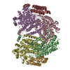
- PDB-7tc8: Cryo-EM structure of methane monooxygenase hydroxylase (by graphene) -
+ Open data
- Basic information
| Entry | Database: PDB / ID: 7tc8 | |||||||||||||||||||||||||||||||||||||||
|---|---|---|---|---|---|---|---|---|---|---|---|---|---|---|---|---|---|---|---|---|---|---|---|---|---|---|---|---|---|---|---|---|---|---|---|---|---|---|---|---|
| Title | Cryo-EM structure of methane monooxygenase hydroxylase (by graphene) | |||||||||||||||||||||||||||||||||||||||
 Components Components |
| |||||||||||||||||||||||||||||||||||||||
 Keywords Keywords | OXIDOREDUCTASE / methane monooxygenase / hydroxylase / cryo-EM / electron transport | |||||||||||||||||||||||||||||||||||||||
| Function / homology |  Function and homology information Function and homology informationmethane metabolic process / methane monooxygenase (soluble) / methane monooxygenase [NAD(P)H] activity / one-carbon metabolic process / metal ion binding Similarity search - Function | |||||||||||||||||||||||||||||||||||||||
| Biological species |  Methylococcus capsulatus (bacteria) Methylococcus capsulatus (bacteria) | |||||||||||||||||||||||||||||||||||||||
| Method | ELECTRON MICROSCOPY / single particle reconstruction / cryo EM / Resolution: 2.4 Å | |||||||||||||||||||||||||||||||||||||||
 Authors Authors | Cho, U.S. / Kim, B.C. | |||||||||||||||||||||||||||||||||||||||
| Funding support |  United States, 3items United States, 3items
| |||||||||||||||||||||||||||||||||||||||
 Citation Citation |  Journal: ACS Nano / Year: 2023 Journal: ACS Nano / Year: 2023Title: Batch Production of High-Quality Graphene Grids for Cryo-EM: Cryo-EM Structure of Soluble Methane Monooxygenase Hydroxylase. Authors: Eungjin Ahn / Byungchul Kim / Soyoung Park / Amanda L Erwin / Suk Hyun Sung / Robert Hovden / Shyamal Mosalaganti / Uhn-Soo Cho /   Abstract: Cryogenic electron microscopy (cryo-EM) has become a widely used tool for determining the protein structure. Despite recent technical advances, sample preparation remains a major bottleneck for ...Cryogenic electron microscopy (cryo-EM) has become a widely used tool for determining the protein structure. Despite recent technical advances, sample preparation remains a major bottleneck for several reasons, including protein denaturation at the air-water interface, the presence of preferred orientations, nonuniform ice layers, etc. Graphene, a two-dimensional allotrope of carbon consisting of a single atomic layer, has recently gained attention as a near-ideal support film for cryo-EM that can overcome these challenges because of its superior properties, including mechanical strength and electrical conductivity. Here, we introduce a reliable, easily implemented, and reproducible method to produce 36 graphene-coated grids within 1.5 days. To demonstrate their practical application, we determined the cryo-EM structure of soluble methane monooxygenase hydroxylase (sMMOH) at resolutions of 2.9 and 2.5 Å using Quantifoil and graphene-coated grids, respectively. We found that the graphene-coated grid has several advantages, including a smaller amount of protein required and avoiding protein denaturation at the air-water interface. By comparing the cryo-EM structure of sMMOH with its crystal structure, we identified subtle yet significant geometrical changes at the nonheme diiron center, which may better indicate the active site configuration of sMMOH in the resting/oxidized state. | |||||||||||||||||||||||||||||||||||||||
| History |
|
- Structure visualization
| Structure viewer | Molecule:  Molmil Molmil Jmol/JSmol Jmol/JSmol |
|---|
- Downloads & links
- Download
| PDBx/mmCIF format |  7tc8.cif.gz 7tc8.cif.gz | 438 KB | Display |  PDBx/mmCIF format PDBx/mmCIF format |
|---|---|---|---|---|
| PDB format |  pdb7tc8.ent.gz pdb7tc8.ent.gz | 364.4 KB | Display |  PDB format PDB format |
| PDBx/mmJSON format |  7tc8.json.gz 7tc8.json.gz | Tree view |  PDBx/mmJSON format PDBx/mmJSON format | |
| Others |  Other downloads Other downloads |
-Validation report
| Summary document |  7tc8_validation.pdf.gz 7tc8_validation.pdf.gz | 894.5 KB | Display |  wwPDB validaton report wwPDB validaton report |
|---|---|---|---|---|
| Full document |  7tc8_full_validation.pdf.gz 7tc8_full_validation.pdf.gz | 933.2 KB | Display | |
| Data in XML |  7tc8_validation.xml.gz 7tc8_validation.xml.gz | 56.7 KB | Display | |
| Data in CIF |  7tc8_validation.cif.gz 7tc8_validation.cif.gz | 90.4 KB | Display | |
| Arichive directory |  https://data.pdbj.org/pub/pdb/validation_reports/tc/7tc8 https://data.pdbj.org/pub/pdb/validation_reports/tc/7tc8 ftp://data.pdbj.org/pub/pdb/validation_reports/tc/7tc8 ftp://data.pdbj.org/pub/pdb/validation_reports/tc/7tc8 | HTTPS FTP |
-Related structure data
| Related structure data |  25805MC  7tc7C M: map data used to model this data C: citing same article ( |
|---|---|
| Similar structure data | Similarity search - Function & homology  F&H Search F&H Search |
- Links
Links
- Assembly
| Deposited unit | 
|
|---|---|
| 1 |
|
- Components
| #1: Protein | Mass: 60717.145 Da / Num. of mol.: 2 / Source method: isolated from a natural source / Source: (natural)  Methylococcus capsulatus (bacteria) / Strain: ATCC 33009 / NCIMB 11132 / Bath Methylococcus capsulatus (bacteria) / Strain: ATCC 33009 / NCIMB 11132 / BathReferences: UniProt: P22869, methane monooxygenase (soluble) #2: Protein | Mass: 45184.660 Da / Num. of mol.: 2 / Source method: isolated from a natural source / Source: (natural)  Methylococcus capsulatus (bacteria) Methylococcus capsulatus (bacteria)References: UniProt: P18798, methane monooxygenase (soluble) #3: Protein | Mass: 19879.732 Da / Num. of mol.: 2 / Source method: isolated from a natural source / Source: (natural)  Methylococcus capsulatus (bacteria) / Strain: ATCC 33009 / NCIMB 11132 / Bath Methylococcus capsulatus (bacteria) / Strain: ATCC 33009 / NCIMB 11132 / BathReferences: UniProt: P11987, methane monooxygenase (soluble) #4: Chemical | ChemComp-FE / Has ligand of interest | Y | Has protein modification | N | |
|---|
-Experimental details
-Experiment
| Experiment | Method: ELECTRON MICROSCOPY |
|---|---|
| EM experiment | Aggregation state: PARTICLE / 3D reconstruction method: single particle reconstruction |
- Sample preparation
| Component | Name: Soluble Methane Monooxygenase Hydroxylase / Type: COMPLEX / Entity ID: #1-#3 / Source: NATURAL |
|---|---|
| Source (natural) | Organism:  Methylococcus capsulatus str. Bath (bacteria) Methylococcus capsulatus str. Bath (bacteria) |
| Buffer solution | pH: 7 / Details: 30 mM HEPES, pH 7, 150 mM NaCl, 1 mM TCEP |
| Specimen | Embedding applied: NO / Shadowing applied: NO / Staining applied: NO / Vitrification applied: YES |
| Specimen support | Grid material: GOLD / Grid mesh size: 300 divisions/in. / Grid type: Quantifoil R1.2/1.3 |
| Vitrification | Instrument: FEI VITROBOT MARK IV / Cryogen name: ETHANE / Humidity: 100 % |
- Electron microscopy imaging
| Experimental equipment |  Model: Titan Krios / Image courtesy: FEI Company |
|---|---|
| Microscopy | Model: FEI TITAN KRIOS |
| Electron gun | Electron source:  FIELD EMISSION GUN / Accelerating voltage: 300 kV / Illumination mode: FLOOD BEAM FIELD EMISSION GUN / Accelerating voltage: 300 kV / Illumination mode: FLOOD BEAM |
| Electron lens | Mode: BRIGHT FIELD / Nominal magnification: 81000 X / Nominal defocus max: 2000 nm / Nominal defocus min: 700 nm / Cs: 2.7 mm / C2 aperture diameter: 50 µm / Alignment procedure: BASIC |
| Specimen holder | Cryogen: NITROGEN / Specimen holder model: FEI TITAN KRIOS AUTOGRID HOLDER |
| Image recording | Electron dose: 51 e/Å2 / Film or detector model: GATAN K3 (6k x 4k) |
- Processing
| Software | Name: PHENIX / Version: 1.18.2_3874: / Classification: refinement | ||||||||||||||||||||||||
|---|---|---|---|---|---|---|---|---|---|---|---|---|---|---|---|---|---|---|---|---|---|---|---|---|---|
| EM software | Name: PHENIX / Category: model refinement | ||||||||||||||||||||||||
| CTF correction | Type: PHASE FLIPPING ONLY | ||||||||||||||||||||||||
| 3D reconstruction | Resolution: 2.4 Å / Resolution method: FSC 0.5 CUT-OFF / Num. of particles: 325000 / Symmetry type: POINT | ||||||||||||||||||||||||
| Refine LS restraints |
|
 Movie
Movie Controller


 PDBj
PDBj


