 Movie
Movie Controller


+ Open data

- Basic information
| Entry | Database: PDB / ID: 7tc5 | ||||||
|---|---|---|---|---|---|---|---|
| Title | All Phe-Azurin variant - F15Y | ||||||
 Components Components | Azurin | ||||||
 Keywords Keywords | ELECTRON TRANSPORT / copper ion binding / cupredoxin / azurin / metal ion binding | ||||||
| Function / homology |  Function and homology information Function and homology informationperiplasmic space / electron transfer activity / oxidoreductase activity / copper ion binding Similarity search - Function | ||||||
| Biological species |   Pseudomonas aeruginosa (bacteria) Pseudomonas aeruginosa (bacteria) | ||||||
| Method |  X-RAY DIFFRACTION / SYNCHROTRON / MOLECULAR REPLACEMENT / Resolution: 1.45 Å X-RAY DIFFRACTION / SYNCHROTRON / MOLECULAR REPLACEMENT / Resolution: 1.45 Å | ||||||
 Authors Authors | Fedoretz-Maxwell, B.P. / Worrall, L.J. / Strynadka, N.C.J. / Warren, J.J. | ||||||
| Funding support |  Canada, 1items Canada, 1items
| ||||||
 Citation Citation | Journal: Inorg.Chem. / Year: 2022 Title: The Impact of Second Coordination Sphere Methionine-Aromatic Interactions in Copper Proteins. Authors: Fedoretz-Maxwell, B.P. / Shin, C.H. / MacNeil, G.A. / Worrall, L.J. / Park, R. / Strynadka, N.C.J. / Walsby, C.J. / Warren, J.J. | ||||||
| History |
|
- Structure visualization
| Structure viewer | Molecule: MolmilJmol/JSmol |
|---|
- Downloads & links
-Download
| PDBx/mmCIF format | 7tc5.cif.gz | 68.9 KB | Display | PDBx/mmCIF format |
|---|---|---|---|---|
| PDB format | pdb7tc5.ent.gz | 48.9 KB | Display | PDB format |
| PDBx/mmJSON format | 7tc5.json.gz | Tree view | PDBx/mmJSON format | |
| Others |  Other downloads Other downloads |
-Validation report
| Summary document | 7tc5_validation.pdf.gz | 2.1 MB | Display | wwPDB validaton report |
|---|---|---|---|---|
| Full document | 7tc5_full_validation.pdf.gz | 2.1 MB | Display | |
| Data in XML | 7tc5_validation.xml.gz | 13.8 KB | Display | |
| Data in CIF | 7tc5_validation.cif.gz | 19.6 KB | Display | |
| Arichive directory | https://data.pdbj.org/pub/pdb/validation_reports/tc/7tc5ftp://data.pdbj.org/pub/pdb/validation_reports/tc/7tc5 | HTTPS FTP |
-Related structure data
| Related structure data |  7tc6C  4azuS S: Starting model for refinement C: citing same article ( |
|---|---|
| Similar structure data |
-Links
 PDBj
PDBj- Assembly
| Deposited unit | 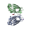
| ||||||||
|---|---|---|---|---|---|---|---|---|---|
| 1 |
| ||||||||
| Unit cell |
|
-Components
| #1: Protein | Mass: 13933.787 Da / Num. of mol.: 2 / Mutation: F15Y, W48F, Y72F, H83Q, Y108F, T124H Source method: isolated from a genetically manipulated source Source: (gene. exp.) Pseudomonas aeruginosa (bacteria) / Gene: azu / Production host: #2: Chemical |   Mass: 63.546 Da / Num. of mol.: 3 / Source method: obtained synthetically / Formula: Cu / Feature type: SUBJECT OF INVESTIGATION Mass: 63.546 Da / Num. of mol.: 3 / Source method: obtained synthetically / Formula: Cu / Feature type: SUBJECT OF INVESTIGATION#3: Chemical | ChemComp-NA / |   Mass: 22.990 Da / Num. of mol.: 1 / Source method: obtained synthetically / Formula: Na Mass: 22.990 Da / Num. of mol.: 1 / Source method: obtained synthetically / Formula: Na#4: Chemical | ChemComp-NO3 / |   Mass: 62.005 Da / Num. of mol.: 1 / Source method: obtained synthetically / Formula: NO3 Mass: 62.005 Da / Num. of mol.: 1 / Source method: obtained synthetically / Formula: NO3#5: Water | ChemComp-HOH / |  Mass: 18.015 Da / Num. of mol.: 212 / Source method: isolated from a natural source / Formula: H2O Mass: 18.015 Da / Num. of mol.: 212 / Source method: isolated from a natural source / Formula: H2OHas ligand of interest | Y | Has protein modification | Y | |
|---|
-Experimental details
-Experiment
| Experiment | Method: X-RAY DIFFRACTION / Number of used crystals: 1 |
|---|
- Sample preparation
| Crystal | Density Matthews: 2.23 Å3/Da / Density % sol: 44.74 % |
|---|---|
| Crystal grow | Temperature: 293 K / Method: vapor diffusion, sitting drop Details: 20 mg/mL protein in 0.1 M NaOAc (pH 5.0), 26.5-29% PEG 4000, 100 mM lithium nitrate, 10 mM copper sulfate, and 100 mM tris (pH 8.0) |
-Data collection
| Diffraction | Mean temperature: 100 K / Serial crystal experiment: N |
|---|---|
| Diffraction source | Source: SYNCHROTRON / Site: CLSI / Beamline: 08B1-1 / Wavelength: 0.97741 Å |
| Detector | Type: DECTRIS PILATUS3 S 6M / Detector: PIXEL / Date: May 1, 2021 |
| Radiation | Protocol: SINGLE WAVELENGTH / Monochromatic (M) / Laue (L): M / Scattering type: x-ray |
| Radiation wavelength | Wavelength: 0.97741 Å / Relative weight: 1 |
| Reflection | Resolution: 1.4→44.6 Å / Num. obs: 45146 / % possible obs: 99.9 % / Redundancy: 6.2 % / CC1/2: 0.999 / Rmerge(I) obs: 0.068 / Net I/σ(I): 11.5 |
| Reflection shell | Resolution: 1.45→1.47 Å / Redundancy: 6.3 % / Rmerge(I) obs: 1.04 / Mean I/σ(I) obs: 1.7 / Num. unique obs: 2184 / CC1/2: 0.683 / % possible all: 99.8 |
- Processing
| Software |
| ||||||||||||||||||||||||||||||||||||||||||||||||||||||||||||
|---|---|---|---|---|---|---|---|---|---|---|---|---|---|---|---|---|---|---|---|---|---|---|---|---|---|---|---|---|---|---|---|---|---|---|---|---|---|---|---|---|---|---|---|---|---|---|---|---|---|---|---|---|---|---|---|---|---|---|---|---|---|
| Refinement | Method to determine structure: MOLECULAR REPLACEMENT Starting model: 4AZU Resolution: 1.45→41.97 Å / Cor.coef. Fo:Fc: 0.972 / Cor.coef. Fo:Fc free: 0.959 / SU B: 1.596 / SU ML: 0.058 / Cross valid method: THROUGHOUT / σ(F): 0 / ESU R: 0.062 / ESU R Free: 0.066 / Stereochemistry target values: MAXIMUM LIKELIHOOD
| ||||||||||||||||||||||||||||||||||||||||||||||||||||||||||||
| Solvent computation | Ion probe radii: 0.8 Å / Shrinkage radii: 0.8 Å / VDW probe radii: 1.2 Å / Solvent model: MASK | ||||||||||||||||||||||||||||||||||||||||||||||||||||||||||||
| Displacement parameters | Biso max: 81.13 Å2 / Biso mean: 23.548 Å2 / Biso min: 12.4 Å2
| ||||||||||||||||||||||||||||||||||||||||||||||||||||||||||||
| Refinement step | Cycle: final / Resolution: 1.45→41.97 Å
| ||||||||||||||||||||||||||||||||||||||||||||||||||||||||||||
| Refine LS restraints |
| ||||||||||||||||||||||||||||||||||||||||||||||||||||||||||||
| LS refinement shell | Resolution: 1.45→1.47 Å / Rfactor Rfree error: 0
|
