 Movie
Movie Controller

[English] 日本語
 Yorodumi
- PDB-7tbc: Crystal structure of Plasmepsin X from Plasmodium falciparum in c... -
+ Open data

- Basic information
| Entry | Database: PDB / ID: 7tbc | ||||||
|---|---|---|---|---|---|---|---|
| Title | Crystal structure of Plasmepsin X from Plasmodium falciparum in complex with WM382 | ||||||
 Components Components | Plasmepsin 10 | ||||||
 Keywords Keywords | HYDROLASE / Aspartyl Protease | ||||||
| Function / homology |  Function and homology information Function and homology informationentry into host cell by a symbiont-containing vacuole / transport vesicle / protein processing / aspartic-type endopeptidase activity Similarity search - Function | ||||||
| Biological species |  | ||||||
| Method |  X-RAY DIFFRACTION / SYNCHROTRON / MOLECULAR REPLACEMENT / Resolution: 2.76 Å X-RAY DIFFRACTION / SYNCHROTRON / MOLECULAR REPLACEMENT / Resolution: 2.76 Å | ||||||
 Authors Authors | Christensen, J.B. / Hodder, A.N. / Scally, S.W. / Cowman, A.F. | ||||||
| Funding support |  United Kingdom, 1items United Kingdom, 1items
| ||||||
 Citation Citation | Journal: Structure / Year: 2022 Title: Basis for drug selectivity of plasmepsin IX and X inhibition in Plasmodium falciparum and vivax. Authors: Hodder, A.N. / Christensen, J. / Scally, S. / Triglia, T. / Ngo, A. / Birkinshaw, R.W. / Bailey, B. / Favuzza, P. / Dietrich, M.H. / Tham, W.H. / Czabotar, P.E. / Lowes, K. / Guo, Z. / ...Authors: Hodder, A.N. / Christensen, J. / Scally, S. / Triglia, T. / Ngo, A. / Birkinshaw, R.W. / Bailey, B. / Favuzza, P. / Dietrich, M.H. / Tham, W.H. / Czabotar, P.E. / Lowes, K. / Guo, Z. / Murgolo, N. / Lera Ruiz, M. / McCauley, J.A. / Sleebs, B.E. / Olsen, D. / Cowman, A.F. | ||||||
| History |
|
- Structure visualization
| Structure viewer | Molecule: MolmilJmol/JSmol |
|---|
- Downloads & links
-Download
| PDBx/mmCIF format | 7tbc.cif.gz | 104.6 KB | Display | PDBx/mmCIF format |
|---|---|---|---|---|
| PDB format | pdb7tbc.ent.gz | 62.6 KB | Display | PDB format |
| PDBx/mmJSON format | 7tbc.json.gz | Tree view | PDBx/mmJSON format | |
| Others |  Other downloads Other downloads |
-Validation report
| Arichive directory | https://data.pdbj.org/pub/pdb/validation_reports/tb/7tbcftp://data.pdbj.org/pub/pdb/validation_reports/tb/7tbc | HTTPS FTP |
|---|
-Related structure data
| Related structure data |  7tbbSC  7tbdC 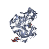 7tbeC S: Starting model for refinement C: citing same article ( |
|---|---|
| Similar structure data |
-Links
 PDBj
PDBj- Assembly
| Deposited unit | 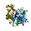
| ||||||||||||
|---|---|---|---|---|---|---|---|---|---|---|---|---|---|
| 1 |
| ||||||||||||
| Unit cell |
|
-Components
-Protein / Sugars , 2 types, 2 molecules A
| #1: Protein | Mass: 42948.523 Da / Num. of mol.: 1 Source method: isolated from a genetically manipulated source Source: (gene. exp.) Production host:   Spodoptera frugiperda (fall armyworm) / References: UniProt: Q2KNW6 Spodoptera frugiperda (fall armyworm) / References: UniProt: Q2KNW6 |
|---|---|
| #3: Sugar | ChemComp-NAG /  Type: D-saccharide, beta linking / Mass: 221.208 Da / Num. of mol.: 1 / Source method: obtained synthetically / Formula: C8H15NO6 Type: D-saccharide, beta linking / Mass: 221.208 Da / Num. of mol.: 1 / Source method: obtained synthetically / Formula: C8H15NO6 |
-Non-polymers , 4 types, 26 molecules 

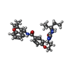

| #2: Chemical | ChemComp-EDO /  Mass: 62.068 Da / Num. of mol.: 15 / Source method: obtained synthetically / Formula: C2H6O2 Mass: 62.068 Da / Num. of mol.: 15 / Source method: obtained synthetically / Formula: C2H6O2#4: Chemical | ChemComp-PG4 / |  Mass: 194.226 Da / Num. of mol.: 1 / Source method: obtained synthetically / Formula: C8H18O5 / Comment: precipitant*YM Mass: 194.226 Da / Num. of mol.: 1 / Source method: obtained synthetically / Formula: C8H18O5 / Comment: precipitant*YM#5: Chemical | ChemComp-I0L / ( |  Mass: 504.621 Da / Num. of mol.: 1 / Source method: obtained synthetically / Formula: C29H36N4O4 / Feature type: SUBJECT OF INVESTIGATION Mass: 504.621 Da / Num. of mol.: 1 / Source method: obtained synthetically / Formula: C29H36N4O4 / Feature type: SUBJECT OF INVESTIGATION#6: Water | ChemComp-HOH / | Mass: 18.015 Da / Num. of mol.: 9 / Source method: isolated from a natural source / Formula: H2O |
|---|
-Details
| Has ligand of interest | Y |
|---|---|
| Has protein modification | Y |
-Experimental details
-Experiment
| Experiment | Method: X-RAY DIFFRACTION / Number of used crystals: 1 |
|---|
- Sample preparation
| Crystal | Density Matthews: 4.75 Å3/Da / Density % sol: 74.12 % |
|---|---|
| Crystal grow | Temperature: 293 K / Method: vapor diffusion, sitting drop / pH: 8.5 Details: 12.5% (v/v) 2-methyl-2,4-pentanediol, 0.1M bicine-tris pH 8.5, 2.85% (v/v) diethylene glycol, 6.35% (v/v) pentaethylene glycol, 12.5% (w/v) polyethylene glycol 1000, 12.5% (w/v) polyethylene ...Details: 12.5% (v/v) 2-methyl-2,4-pentanediol, 0.1M bicine-tris pH 8.5, 2.85% (v/v) diethylene glycol, 6.35% (v/v) pentaethylene glycol, 12.5% (w/v) polyethylene glycol 1000, 12.5% (w/v) polyethylene glycol 3350, 5.2% (v/v) tetraethylene glycol, 4.1% (v/v) triethylene glycol |
-Data collection
| Diffraction | Mean temperature: 100 K / Serial crystal experiment: N |
|---|---|
| Diffraction source | Source: SYNCHROTRON / Site: Australian Synchrotron  / Beamline: MX2 / Wavelength: 0.95373 Å / Beamline: MX2 / Wavelength: 0.95373 Å |
| Detector | Type: DECTRIS EIGER X 16M / Detector: PIXEL / Date: Oct 15, 2020 |
| Radiation | Protocol: SINGLE WAVELENGTH / Monochromatic (M) / Laue (L): M / Scattering type: x-ray |
| Radiation wavelength | Wavelength: 0.95373 Å / Relative weight: 1 |
| Reflection | Resolution: 2.76→49.63 Å / Num. obs: 21289 / % possible obs: 99 % / Redundancy: 22.6 % / Biso Wilson estimate: 96.22 Å2 / CC1/2: 0.999 / Rpim(I) all: 0.03 / Net I/σ(I): 12.5 |
| Reflection shell | Resolution: 2.76→2.91 Å / Redundancy: 6.5 % / Num. unique obs: 2860 / CC1/2: 0.936 / Rpim(I) all: 0.496 / % possible all: 93.6 |
- Processing
| Software |
| |||||||||||||||||||||||||||||||||||||||||||||||||||||||||||||||
|---|---|---|---|---|---|---|---|---|---|---|---|---|---|---|---|---|---|---|---|---|---|---|---|---|---|---|---|---|---|---|---|---|---|---|---|---|---|---|---|---|---|---|---|---|---|---|---|---|---|---|---|---|---|---|---|---|---|---|---|---|---|---|---|---|
| Refinement | Method to determine structure: MOLECULAR REPLACEMENT Starting model: 7TBB Resolution: 2.76→49.62 Å / SU ML: 0.3746 / Cross valid method: FREE R-VALUE / σ(F): 1.34 / Phase error: 27.7843 Stereochemistry target values: GeoStd + Monomer Library + CDL v1.2
| |||||||||||||||||||||||||||||||||||||||||||||||||||||||||||||||
| Solvent computation | Shrinkage radii: 0.9 Å / VDW probe radii: 1.11 Å / Solvent model: FLAT BULK SOLVENT MODEL | |||||||||||||||||||||||||||||||||||||||||||||||||||||||||||||||
| Displacement parameters | Biso mean: 91.48 Å2 | |||||||||||||||||||||||||||||||||||||||||||||||||||||||||||||||
| Refinement step | Cycle: LAST / Resolution: 2.76→49.62 Å
| |||||||||||||||||||||||||||||||||||||||||||||||||||||||||||||||
| Refine LS restraints |
| |||||||||||||||||||||||||||||||||||||||||||||||||||||||||||||||
| LS refinement shell |
|
