 Movie
Movie Controller


+ Open data

- Basic information
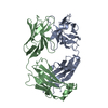
| Entry | Database: PDB / ID: 7t97 | ||||||
|---|---|---|---|---|---|---|---|
| Title | Crystal structure of engineered CYS-CYS fab dimer CH1-207 (HC4) | ||||||
 Components Components |
| ||||||
 Keywords Keywords | IMMUNE SYSTEM | ||||||
| Function / homology | Immunoglobulins / Immunoglobulin-like / Sandwich / Mainly Beta Function and homology information Function and homology information | ||||||
| Biological species |  Homo sapiens (human) Homo sapiens (human) | ||||||
| Method |  X-RAY DIFFRACTION / SYNCHROTRON / MOLECULAR REPLACEMENT / Resolution: 2.144 Å X-RAY DIFFRACTION / SYNCHROTRON / MOLECULAR REPLACEMENT / Resolution: 2.144 Å | ||||||
 Authors Authors | Harris, S.F. / Boenig, G.D.L. | ||||||
| Funding support | 1items
| ||||||
 Citation Citation | Journal: Comput Struct Biotechnol J / Year: 2022 Title: Antibody interfaces revealed through structural mining. Authors: Yin, Y. / Romei, M.G. / Sankar, K. / Pal, L.R. / Hoi, K.H. / Yang, Y. / Leonard, B. / De Leon Boenig, G. / Kumar, N. / Matsumoto, M. / Payandeh, J. / Harris, S.F. / Moult, J. / Lazar, G.A. | ||||||
| History |
|
- Structure visualization
| Structure viewer | Molecule: MolmilJmol/JSmol |
|---|
- Downloads & links
-Download
| PDBx/mmCIF format | 7t97.cif.gz | 347 KB | Display | PDBx/mmCIF format |
|---|---|---|---|---|
| PDB format | pdb7t97.ent.gz | 281.4 KB | Display | PDB format |
| PDBx/mmJSON format | 7t97.json.gz | Tree view | PDBx/mmJSON format | |
| Others |  Other downloads Other downloads |
-Validation report
| Arichive directory | https://data.pdbj.org/pub/pdb/validation_reports/t9/7t97ftp://data.pdbj.org/pub/pdb/validation_reports/t9/7t97 | HTTPS FTP |
|---|
-Related structure data

-Links
 PDBj
PDBj

- Assembly
| Deposited unit | 
| ||||||||
|---|---|---|---|---|---|---|---|---|---|
| 1 | 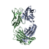
| ||||||||
| 2 | 
| ||||||||
| 3 | 
| ||||||||
| 4 | 
| ||||||||
| Unit cell |
|
-Components
| #1: Antibody | Mass: 24391.359 Da / Num. of mol.: 4 Source method: isolated from a genetically manipulated source Source: (gene. exp.) Homo sapiens (human) / Production host:  #2: Antibody | Mass: 23466.031 Da / Num. of mol.: 4 Source method: isolated from a genetically manipulated source Source: (gene. exp.) Homo sapiens (human) / Production host: #3: Water | ChemComp-HOH / |  Mass: 18.015 Da / Num. of mol.: 740 / Source method: isolated from a natural source / Formula: H2O Mass: 18.015 Da / Num. of mol.: 740 / Source method: isolated from a natural source / Formula: H2OHas protein modification | Y | |
|---|
-Experimental details
-Experiment
| Experiment | Method: X-RAY DIFFRACTION / Number of used crystals: 1 |
|---|
- Sample preparation
| Crystal | Density Matthews: 3.31 Å3/Da / Density % sol: 62.81 % |
|---|---|
| Crystal grow | Temperature: 293 K / Method: vapor diffusion, sitting drop / Details: 0.8 M sodium succinate |
-Data collection
| Diffraction | Mean temperature: 100 K / Serial crystal experiment: N | ||||||||||||||||||||||||||||||
|---|---|---|---|---|---|---|---|---|---|---|---|---|---|---|---|---|---|---|---|---|---|---|---|---|---|---|---|---|---|---|---|
| Diffraction source | Source: SYNCHROTRON / Site: ALS  / Beamline: 5.0.2 / Wavelength: 0.97949 Å / Beamline: 5.0.2 / Wavelength: 0.97949 Å | ||||||||||||||||||||||||||||||
| Detector | Type: DECTRIS PILATUS 6M / Detector: PIXEL / Date: Apr 13, 2021 | ||||||||||||||||||||||||||||||
| Radiation | Protocol: SINGLE WAVELENGTH / Monochromatic (M) / Laue (L): M / Scattering type: x-ray | ||||||||||||||||||||||||||||||
| Radiation wavelength | Wavelength: 0.97949 Å / Relative weight: 1 | ||||||||||||||||||||||||||||||
| Reflection | Resolution: 2.144→75.69 Å / Num. obs: 90661 / % possible obs: 99.8 % / Redundancy: 3.5 % / Biso Wilson estimate: 36.61 Å2 / CC1/2: 0.995 / Rmerge(I) obs: 0.111 / Rpim(I) all: 0.07 / Rrim(I) all: 0.131 / Net I/σ(I): 9.2 | ||||||||||||||||||||||||||||||
| Reflection shell | Diffraction-ID: 1
|
- Processing
| Software |
| ||||||||||||||||||||||||||||||||||||||||||||||||||||||||||||||||||||||||||||||||||||||||||||||||||||||||||||
|---|---|---|---|---|---|---|---|---|---|---|---|---|---|---|---|---|---|---|---|---|---|---|---|---|---|---|---|---|---|---|---|---|---|---|---|---|---|---|---|---|---|---|---|---|---|---|---|---|---|---|---|---|---|---|---|---|---|---|---|---|---|---|---|---|---|---|---|---|---|---|---|---|---|---|---|---|---|---|---|---|---|---|---|---|---|---|---|---|---|---|---|---|---|---|---|---|---|---|---|---|---|---|---|---|---|---|---|---|---|
| Refinement | Method to determine structure: MOLECULAR REPLACEMENT / Resolution: 2.144→75.69 Å / Cor.coef. Fo:Fc: 0.932 / Cor.coef. Fo:Fc free: 0.908 / SU R Cruickshank DPI: 0.3 / Cross valid method: THROUGHOUT / σ(F): 0 / SU R Blow DPI: 0.299 / SU Rfree Blow DPI: 0.215 / SU Rfree Cruickshank DPI: 0.218
| ||||||||||||||||||||||||||||||||||||||||||||||||||||||||||||||||||||||||||||||||||||||||||||||||||||||||||||
| Displacement parameters | Biso max: 217.71 Å2 / Biso mean: 35.98 Å2 / Biso min: 15.09 Å2
| ||||||||||||||||||||||||||||||||||||||||||||||||||||||||||||||||||||||||||||||||||||||||||||||||||||||||||||
| Refine analyze | Luzzati coordinate error obs: 0.32 Å | ||||||||||||||||||||||||||||||||||||||||||||||||||||||||||||||||||||||||||||||||||||||||||||||||||||||||||||
| Refinement step | Cycle: final / Resolution: 2.144→75.69 Å
| ||||||||||||||||||||||||||||||||||||||||||||||||||||||||||||||||||||||||||||||||||||||||||||||||||||||||||||
| Refine LS restraints |
| ||||||||||||||||||||||||||||||||||||||||||||||||||||||||||||||||||||||||||||||||||||||||||||||||||||||||||||
| LS refinement shell | Resolution: 2.144→2.22 Å / Rfactor Rfree error: 0
|
