Mass: 18.015 Da / Num. of mol.: 19 / Source method: isolated from a natural source / Formula: H2O
Compound details
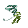
The crystallized sequence is: ...The crystallized sequence is: GSHMSAVNNAYDALIIEARKGNTQPALSWFALKSALSNNQIADWLQIALWAGQDKQVITVYNRYRHQQLPARGYAAVAVA YRNLQQWQNSLTLWQKALSLEPQNKDYQRGQILTLADAGHYDTALVKLKQLNSGAPDKANLLAEAYIYKLAGRHQDELRA MTESLPENASTQQYPTEYVQALRNNQLAAAIDDANLTPDIRADIHAELVRLSFMPTRSESERYAIADRALAQYAALEILW HDNPDRTAQYQRIQVDHLGALLTRDRYKDVISHYQRLKKTGQIIPPWGQYWVASAYLKDHQPKKAQSIMTELFYHKETIA PDLSDEELADLFYSHLESEN. As per the authors, the protein must have cleaved and only the C-terminal fragment crystallized. They do not know exactly at which residue the cleavage occurred.
Has ligand of interest
N
-
Experimental details
-
Experiment
Experiment
Method: X-RAY DIFFRACTION / Number of used crystals: 1
-
Sample preparation
Crystal
Density Matthews: 2.85 Å3/Da / Density % sol: 61.95 %
Crystal grow
Temperature: 300 K / Method: vapor diffusion, hanging drop Details: 15% w/v PEG 8000, 0.1 M Tris pH 7.1, 200 mM MgCl2, and 3% w/v cadaverine
-
Data collection
Diffraction
Mean temperature: 100 K / Serial crystal experiment: N
Method to determine structure: SAD / Resolution: 2.85→20.001 Å / Cor.coef. Fo:Fc: 0.91 / Cor.coef. Fo:Fc free: 0.878 / SU B: 41.677 / SU ML: 0.344 / Cross valid method: FREE R-VALUE / ESU R Free: 0.414 Details: Hydrogens have been added in their riding positions
Rfactor
Num. reflection
% reflection
Rfree
0.2643
499
5.967 %
Rwork
0.2347
7863
-
all
0.237
-
-
obs
-
8362
92.469 %
Solvent computation
Ion probe radii: 0.8 Å / Shrinkage radii: 0.8 Å / VDW probe radii: 1.2 Å / Solvent model: MASK BULK SOLVENT
Displacement parameters
Biso mean: 49.606 Å2
Baniso -1
Baniso -2
Baniso -3
1-
-1.598 Å2
-0.799 Å2
-0 Å2
2-
-
-1.598 Å2
0 Å2
3-
-
-
5.184 Å2
Refinement step
Cycle: LAST / Resolution: 2.85→20.001 Å
Protein
Nucleic acid
Ligand
Solvent
Total
Num. atoms
2125
0
3
19
2147
Refine LS restraints
Refine-ID
Type
Dev ideal
Dev ideal target
Number
X-RAY DIFFRACTION
r_bond_refined_d
0.007
0.013
2205
X-RAY DIFFRACTION
r_bond_other_d
0.001
0.017
2034
X-RAY DIFFRACTION
r_angle_refined_deg
1.424
1.647
3000
X-RAY DIFFRACTION
r_angle_other_deg
1.231
1.582
4667
X-RAY DIFFRACTION
r_dihedral_angle_1_deg
5.795
5
262
X-RAY DIFFRACTION
r_dihedral_angle_2_deg
32.137
21.212
132
X-RAY DIFFRACTION
r_dihedral_angle_3_deg
19.091
15
363
X-RAY DIFFRACTION
r_dihedral_angle_4_deg
15.061
15
19
X-RAY DIFFRACTION
r_chiral_restr
0.06
0.2
286
X-RAY DIFFRACTION
r_gen_planes_refined
0.006
0.02
2499
X-RAY DIFFRACTION
r_gen_planes_other
0.001
0.02
535
X-RAY DIFFRACTION
r_nbd_refined
0.226
0.2
523
X-RAY DIFFRACTION
r_symmetry_nbd_other
0.2
0.2
1852
X-RAY DIFFRACTION
r_nbtor_refined
0.168
0.2
1051
X-RAY DIFFRACTION
r_symmetry_nbtor_other
0.084
0.2
1038
X-RAY DIFFRACTION
r_xyhbond_nbd_refined
0.175
0.2
53
X-RAY DIFFRACTION
r_symmetry_nbd_refined
0.204
0.2
20
X-RAY DIFFRACTION
r_nbd_other
0.291
0.2
60
X-RAY DIFFRACTION
r_symmetry_xyhbond_nbd_refined
0.201
0.2
2
X-RAY DIFFRACTION
r_mcbond_it
1.395
2.666
1048
X-RAY DIFFRACTION
r_mcbond_other
1.391
2.663
1047
X-RAY DIFFRACTION
r_mcangle_it
2.475
3.987
1310
X-RAY DIFFRACTION
r_mcangle_other
2.474
3.991
1311
X-RAY DIFFRACTION
r_scbond_it
1.137
2.772
1156
X-RAY DIFFRACTION
r_scbond_other
1.137
2.775
1157
X-RAY DIFFRACTION
r_scangle_it
2.053
4.109
1689
X-RAY DIFFRACTION
r_scangle_other
2.053
4.111
1690
X-RAY DIFFRACTION
r_lrange_it
4.794
29.988
2544
X-RAY DIFFRACTION
r_lrange_other
4.794
30.011
2545
X-RAY DIFFRACTION
r_ncsr_local_group_1
0.131
0.05
3782
Refine LS restraints NCS
Ens-ID
Dom-ID
Auth asym-ID
Refine-ID
Type
Rms dev position (Å)
Weight position
1
1
AAA
X-RAY DIFFRACTION
Localncs
0.13127
0.05007
1
2
BBB
X-RAY DIFFRACTION
Localncs
0.13127
0.05007
LS refinement shell
Refine-ID: X-RAY DIFFRACTION / Total num. of bins used: 20
Resolution (Å)
Rfactor Rfree
Num. reflection Rfree
Rfactor Rwork
Num. reflection Rwork
Rfactor all
Num. reflection all
Fsc free
Fsc work
% reflection obs (%)
WRfactor Rwork
2.85-2.922
0.207
11
0.277
236
0.274
666
0.852
0.767
37.0871
0.259
2.922-3.001
0.341
20
0.318
381
0.319
624
0.857
0.788
64.2628
0.259
3.001-3.086
0.33
38
0.344
588
0.343
627
0.741
0.803
99.8405
0.258
3.086-3.178
0.255
36
0.319
561
0.315
598
0.83
0.848
99.8328
0.231
3.178-3.28
0.329
37
0.276
542
0.279
580
0.825
0.877
99.8276
0.207
3.28-3.392
0.287
34
0.265
535
0.266
569
0.875
0.885
100
0.2
3.392-3.516
0.282
35
0.256
514
0.257
549
0.884
0.887
100
0.219
3.516-3.655
0.311
30
0.252
492
0.255
522
0.877
0.899
100
0.196
3.655-3.811
0.268
32
0.226
480
0.229
512
0.904
0.914
100
0.186
3.811-3.99
0.309
32
0.223
443
0.229
475
0.882
0.919
100
0.186
3.99-4.197
0.237
23
0.215
435
0.216
459
0.938
0.933
99.7821
0.18
4.197-4.439
0.309
28
0.181
416
0.188
444
0.908
0.952
100
0.156
4.439-4.729
0.212
25
0.179
394
0.181
419
0.955
0.961
100
0.159
4.729-5.084
0.17
26
0.212
360
0.209
386
0.973
0.948
100
0.199
5.084-5.534
0.32
24
0.233
342
0.239
366
0.922
0.937
100
0.209
5.534-6.129
0.203
15
0.215
305
0.214
320
0.958
0.937
100
0.192
6.129-6.969
0.172
17
0.207
280
0.205
297
0.957
0.949
100
0.195
6.969-8.287
0.247
15
0.184
226
0.188
241
0.962
0.965
100
0.189
8.287-10.827
0.217
13
0.18
195
0.182
209
0.968
0.978
99.5215
0.182
10.827-20.001
0.335
8
0.264
137
0.268
147
0.876
0.946
98.6395
0.283
Refinement TLS params.
Method: refined / Refine-ID: X-RAY DIFFRACTION
ID
L11 (°2)
L12 (°2)
L13 (°2)
L22 (°2)
L23 (°2)
L33 (°2)
S11 (Å °)
S12 (Å °)
S13 (Å °)
S21 (Å °)
S22 (Å °)
S23 (Å °)
S31 (Å °)
S32 (Å °)
S33 (Å °)
T11 (Å2)
T12 (Å2)
T13 (Å2)
T22 (Å2)
T23 (Å2)
T33 (Å2)
Origin x (Å)
Origin y (Å)
Origin z (Å)
1
1.3353
0.2058
1.2802
0.2438
-0.0867
1.9893
0.0751
-0.1602
0.0225
-0.0637
-0.0022
0.0607
-0.1688
-0.1733
-0.0729
0.391
0.1359
0.0131
0.4226
0.0369
0.02
4.421
34.288
52.696
2
2.6314
-0.322
1.7147
2.2587
-0.2424
3.2472
0.0972
-0.2396
0.2161
0.067
-0.134
0.2208
-0.1371
-0.134
0.0368
0.2118
0.0415
0.0814
0.3467
-0.0033
0.0816
2.625
34.163
82.92
Refinement TLS group
ID
Refine-ID
Refine TLS-ID
Selection
Auth asym-ID
Auth seq-ID
1
X-RAY DIFFRACTION
1
ALL
AAA
224 - 359
2
X-RAY DIFFRACTION
2
ALL
BBB
220 - 343
+
About Yorodumi
-
News
-
Feb 9, 2022. New format data for meta-information of EMDB entries
New format data for meta-information of EMDB entries
Version 3 of the EMDB header file is now the official format.
The previous official version 1.9 will be removed from the archive.
In the structure databanks used in Yorodumi, some data are registered as the other names, "COVID-19 virus" and "2019-nCoV". Here are the details of the virus and the list of structure data.
Jan 31, 2019. EMDB accession codes are about to change! (news from PDBe EMDB page)
EMDB accession codes are about to change! (news from PDBe EMDB page)
The allocation of 4 digits for EMDB accession codes will soon come to an end. Whilst these codes will remain in use, new EMDB accession codes will include an additional digit and will expand incrementally as the available range of codes is exhausted. The current 4-digit format prefixed with “EMD-” (i.e. EMD-XXXX) will advance to a 5-digit format (i.e. EMD-XXXXX), and so on. It is currently estimated that the 4-digit codes will be depleted around Spring 2019, at which point the 5-digit format will come into force.
The EM Navigator/Yorodumi systems omit the EMD- prefix.
Related info.:Q: What is EMD? / ID/Accession-code notation in Yorodumi/EM Navigator
Yorodumi is a browser for structure data from EMDB, PDB, SASBDB, etc.
This page is also the successor to EM Navigator detail page, and also detail information page/front-end page for Omokage search.
The word "yorodu" (or yorozu) is an old Japanese word meaning "ten thousand". "mi" (miru) is to see.
Related info.:EMDB / PDB / SASBDB / Comparison of 3 databanks / Yorodumi Search / Aug 31, 2016. New EM Navigator & Yorodumi / Yorodumi Papers / Jmol/JSmol / Function and homology information / Changes in new EM Navigator and Yorodumi
 Movie
Movie Controller


 Open data
 Basic information
 Components
 Keywords
 STRUCTURAL PROTEIN / PNAG binding module / tetra-trico repeat (TPR) domain / PNAG secretion
 Function and homology information
 Authors
 Canada, 1items
  Citation
 Structure visualization
 Downloads & links
 Other downloads
 PDBj
PDBj


 Assembly

 Mass: 24.305 Da / Num. of mol.: 2 / Source method: obtained synthetically / Formula: Mg
 Mass: 35.453 Da / Num. of mol.: 1 / Source method: obtained synthetically / Formula: Cl
 Mass: 18.015 Da / Num. of mol.: 19 / Source method: isolated from a natural source / Formula: H2O
 Sample preparation
 / Beamline: X29A / Wavelength: 0.979 Å
 Processing