 Movie
Movie Controller


+ Open data

- Basic information
| Entry | Database: PDB / ID: 7t80 | ||||||
|---|---|---|---|---|---|---|---|
| Title | Crystal Structure of Mouse Cadherin-23 EC18-19 | ||||||
 Components Components | Cadherin-23 | ||||||
 Keywords Keywords | CELL ADHESION / HEARING / MECHANOTRANSDUCTION / ADHESION / CALCIUM-BINDING PROTEIN | ||||||
| Function / homology |  Function and homology information Function and homology informationequilibrioception / sensory perception of light stimulus / cochlear hair cell ribbon synapse / stereocilium tip / inner ear receptor cell stereocilium organization / photoreceptor ribbon synapse / kinocilium / inner ear auditory receptor cell differentiation / calcium-dependent cell-cell adhesion via plasma membrane cell adhesion molecules / stereocilium ...equilibrioception / sensory perception of light stimulus / cochlear hair cell ribbon synapse / stereocilium tip / inner ear receptor cell stereocilium organization / photoreceptor ribbon synapse / kinocilium / inner ear auditory receptor cell differentiation / calcium-dependent cell-cell adhesion via plasma membrane cell adhesion molecules / stereocilium / photoreceptor cell maintenance / auditory receptor cell stereocilium organization / inner ear morphogenesis / homophilic cell adhesion via plasma membrane adhesion molecules / inner ear development / cochlea development / regulation of cytosolic calcium ion concentration / photoreceptor inner segment / locomotory behavior / sensory perception of sound / calcium ion transport / cell adhesion / calcium ion binding / synapse / centrosome / plasma membrane Similarity search - Function | ||||||
| Biological species |  | ||||||
| Method |  X-RAY DIFFRACTION / SYNCHROTRON / MOLECULAR REPLACEMENT / Resolution: 2.301 Å X-RAY DIFFRACTION / SYNCHROTRON / MOLECULAR REPLACEMENT / Resolution: 2.301 Å | ||||||
 Authors Authors | Harrison-Rawn, T. / Sotomayor, M. | ||||||
| Funding support |  United States, 1items United States, 1items
| ||||||
 Citation Citation | Journal: to be published Title: Crystal Structure of Mouse Cadherin-23 EC18-19 Authors: Harrison-Rawn, T. / Sotomayor, M. | ||||||
| History |
|
- Structure visualization
| Structure viewer | Molecule: MolmilJmol/JSmol |
|---|
- Downloads & links
-Download
| PDBx/mmCIF format | 7t80.cif.gz | 103.8 KB | Display | PDBx/mmCIF format |
|---|---|---|---|---|
| PDB format | pdb7t80.ent.gz | 74.1 KB | Display | PDB format |
| PDBx/mmJSON format | 7t80.json.gz | Tree view | PDBx/mmJSON format | |
| Others |  Other downloads Other downloads |
-Validation report
| Summary document | 7t80_validation.pdf.gz | 2.7 MB | Display | wwPDB validaton report |
|---|---|---|---|---|
| Full document | 7t80_full_validation.pdf.gz | 2.7 MB | Display | |
| Data in XML | 7t80_validation.xml.gz | 17.1 KB | Display | |
| Data in CIF | 7t80_validation.cif.gz | 23.4 KB | Display | |
| Arichive directory | https://data.pdbj.org/pub/pdb/validation_reports/t8/7t80ftp://data.pdbj.org/pub/pdb/validation_reports/t8/7t80 | HTTPS FTP |
-Related structure data


-Links
 PDBj
PDBj
- Assembly
| Deposited unit | 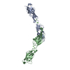
| ||||||||
|---|---|---|---|---|---|---|---|---|---|
| 1 | 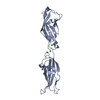
| ||||||||
| 2 | 
| ||||||||
| Unit cell |
|
-Components
| #1: Protein | Mass: 24866.822 Da / Num. of mol.: 2 Source method: isolated from a genetically manipulated source Source: (gene. exp.)  #2: Chemical | ChemComp-CA /   Mass: 40.078 Da / Num. of mol.: 6 / Source method: obtained synthetically / Formula: Ca / Feature type: SUBJECT OF INVESTIGATION Mass: 40.078 Da / Num. of mol.: 6 / Source method: obtained synthetically / Formula: Ca / Feature type: SUBJECT OF INVESTIGATION#3: Chemical |   Mass: 35.453 Da / Num. of mol.: 2 / Source method: obtained synthetically / Formula: Cl Mass: 35.453 Da / Num. of mol.: 2 / Source method: obtained synthetically / Formula: Cl#4: Water | ChemComp-HOH / |  Mass: 18.015 Da / Num. of mol.: 68 / Source method: isolated from a natural source / Formula: H2O Mass: 18.015 Da / Num. of mol.: 68 / Source method: isolated from a natural source / Formula: H2OHas ligand of interest | Y | |
|---|
-Experimental details
-Experiment
| Experiment | Method: X-RAY DIFFRACTION / Number of used crystals: 1 |
|---|
- Sample preparation
| Crystal | Density Matthews: 3.38 Å3/Da / Density % sol: 63.65 % |
|---|---|
| Crystal grow | Temperature: 277 K / Method: vapor diffusion, sitting drop / pH: 8.5 Details: 0.2 M (NH4)2SO4 25% w/v PEG 3350 0.1 M Tris-HCl 8.5 pH |
-Data collection
| Diffraction | Mean temperature: 100 K / Serial crystal experiment: N |
|---|---|
| Diffraction source | Source: SYNCHROTRON / Site: APS / Beamline: 24-ID-C / Wavelength: 0.97918 Å |
| Detector | Type: DECTRIS EIGER2 X 16M / Detector: PIXEL / Date: Jul 29, 2021 |
| Radiation | Protocol: SINGLE WAVELENGTH / Monochromatic (M) / Laue (L): M / Scattering type: x-ray |
| Radiation wavelength | Wavelength: 0.97918 Å / Relative weight: 1 |
| Reflection | Resolution: 2.3→67.106 Å / Num. obs: 30864 / % possible obs: 100 % / Redundancy: 17.6 % / CC1/2: 0.999 / Net I/σ(I): 21.28 |
| Reflection shell | Resolution: 2.3→2.34 Å / Redundancy: 13.4 % / Mean I/σ(I) obs: 2.7 / Num. unique obs: 1495 / CC1/2: 0.706 / % possible all: 99.9 |
- Processing
| Software |
| |||||||||||||||||||||||||||||||||||||||||||||||||||||||||||||||||||||||||||||||||||||||||||||||||||||||||||||||||||||||||||||||||||||||||||||||||||||||||||||||||||||||||||||||||||||||||||||||||||||||||||||||||||||||||||||||||||||||
|---|---|---|---|---|---|---|---|---|---|---|---|---|---|---|---|---|---|---|---|---|---|---|---|---|---|---|---|---|---|---|---|---|---|---|---|---|---|---|---|---|---|---|---|---|---|---|---|---|---|---|---|---|---|---|---|---|---|---|---|---|---|---|---|---|---|---|---|---|---|---|---|---|---|---|---|---|---|---|---|---|---|---|---|---|---|---|---|---|---|---|---|---|---|---|---|---|---|---|---|---|---|---|---|---|---|---|---|---|---|---|---|---|---|---|---|---|---|---|---|---|---|---|---|---|---|---|---|---|---|---|---|---|---|---|---|---|---|---|---|---|---|---|---|---|---|---|---|---|---|---|---|---|---|---|---|---|---|---|---|---|---|---|---|---|---|---|---|---|---|---|---|---|---|---|---|---|---|---|---|---|---|---|---|---|---|---|---|---|---|---|---|---|---|---|---|---|---|---|---|---|---|---|---|---|---|---|---|---|---|---|---|---|---|---|---|---|---|---|---|---|---|---|---|---|---|---|---|---|---|---|---|---|
| Refinement | Method to determine structure: MOLECULAR REPLACEMENT Starting model: 5WJM, 5TFK Resolution: 2.301→67.106 Å / Cor.coef. Fo:Fc: 0.957 / Cor.coef. Fo:Fc free: 0.939 / SU B: 6.246 / SU ML: 0.144 / Cross valid method: FREE R-VALUE / ESU R: 0.215 / ESU R Free: 0.184 Details: Hydrogens have been added in their riding positions
| |||||||||||||||||||||||||||||||||||||||||||||||||||||||||||||||||||||||||||||||||||||||||||||||||||||||||||||||||||||||||||||||||||||||||||||||||||||||||||||||||||||||||||||||||||||||||||||||||||||||||||||||||||||||||||||||||||||||
| Solvent computation | Ion probe radii: 0.8 Å / Shrinkage radii: 0.8 Å / VDW probe radii: 1.2 Å / Solvent model: MASK BULK SOLVENT | |||||||||||||||||||||||||||||||||||||||||||||||||||||||||||||||||||||||||||||||||||||||||||||||||||||||||||||||||||||||||||||||||||||||||||||||||||||||||||||||||||||||||||||||||||||||||||||||||||||||||||||||||||||||||||||||||||||||
| Displacement parameters | Biso mean: 58.004 Å2
| |||||||||||||||||||||||||||||||||||||||||||||||||||||||||||||||||||||||||||||||||||||||||||||||||||||||||||||||||||||||||||||||||||||||||||||||||||||||||||||||||||||||||||||||||||||||||||||||||||||||||||||||||||||||||||||||||||||||
| Refinement step | Cycle: LAST / Resolution: 2.301→67.106 Å
| |||||||||||||||||||||||||||||||||||||||||||||||||||||||||||||||||||||||||||||||||||||||||||||||||||||||||||||||||||||||||||||||||||||||||||||||||||||||||||||||||||||||||||||||||||||||||||||||||||||||||||||||||||||||||||||||||||||||
| Refine LS restraints |
| |||||||||||||||||||||||||||||||||||||||||||||||||||||||||||||||||||||||||||||||||||||||||||||||||||||||||||||||||||||||||||||||||||||||||||||||||||||||||||||||||||||||||||||||||||||||||||||||||||||||||||||||||||||||||||||||||||||||
| LS refinement shell | Refine-ID: X-RAY DIFFRACTION / Total num. of bins used: 20
|
