- PDB-7t7z: The crystal structure of family 8 carbohydrate-binding module fro... -
+
Open data
ID or keywords:
Loading...
-
Basic information
Entry
Database: PDB / ID: 7t7z
Title
The crystal structure of family 8 carbohydrate-binding module from Dictyostelium discoideum
Components
Endoglucanase
Keywords
SUGAR BINDING PROTEIN / Carbohydrate-binding protein / ligand specificity / cellulose / CAZymes
Function / homology
Function and homology information
spore germination / cellulose binding / cellulase / cellulase activity / sporulation resulting in formation of a cellular spore / cellulose catabolic process / extracellular region Similarity search - Function
Glycosyl hydrolases family 9 (GH9) active site signature 1. / Glycoside hydrolase family 9, His active site / Glycosyl hydrolases family 9 (GH9) active site signature 2. / Galactose-binding lectin / Glycosyl hydrolases family 9, Asp/Glu active sites / Glycosyl hydrolases family 9 (GH9) active site signature 3. / Glycoside hydrolase family 9 / Glycosyl hydrolase family 9 / Six-hairpin glycosidase-like superfamily / Six-hairpin glycosidase superfamily ...Glycosyl hydrolases family 9 (GH9) active site signature 1. / Glycoside hydrolase family 9, His active site / Glycosyl hydrolases family 9 (GH9) active site signature 2. / Galactose-binding lectin / Glycosyl hydrolases family 9, Asp/Glu active sites / Glycosyl hydrolases family 9 (GH9) active site signature 3. / Glycoside hydrolase family 9 / Glycosyl hydrolase family 9 / Six-hairpin glycosidase-like superfamily / Six-hairpin glycosidase superfamily / Jelly Rolls / Sandwich / Mainly Beta Similarity search - Domain/homology
In the structure databanks used in Yorodumi, some data are registered as the other names, "COVID-19 virus" and "2019-nCoV". Here are the details of the virus and the list of structure data.
Jan 31, 2019. EMDB accession codes are about to change! (news from PDBe EMDB page)
EMDB accession codes are about to change! (news from PDBe EMDB page)
The allocation of 4 digits for EMDB accession codes will soon come to an end. Whilst these codes will remain in use, new EMDB accession codes will include an additional digit and will expand incrementally as the available range of codes is exhausted. The current 4-digit format prefixed with “EMD-” (i.e. EMD-XXXX) will advance to a 5-digit format (i.e. EMD-XXXXX), and so on. It is currently estimated that the 4-digit codes will be depleted around Spring 2019, at which point the 5-digit format will come into force.
The EM Navigator/Yorodumi systems omit the EMD- prefix.
Related info.:Q: What is EMD? / ID/Accession-code notation in Yorodumi/EM Navigator
Yorodumi is a browser for structure data from EMDB, PDB, SASBDB, etc.
This page is also the successor to EM Navigator detail page, and also detail information page/front-end page for Omokage search.
The word "yorodu" (or yorozu) is an old Japanese word meaning "ten thousand". "mi" (miru) is to see.
Related info.:EMDB / PDB / SASBDB / Comparison of 3 databanks / Yorodumi Search / Aug 31, 2016. New EM Navigator & Yorodumi / Yorodumi Papers / Jmol/JSmol / Function and homology information / Changes in new EM Navigator and Yorodumi
 Movie
Movie Controller

 Yorodumi
 Open data
 Basic information
 Components
 Keywords
 Function and homology information
 X-RAY DIFFRACTION /
  Authors
 Brazil, 3items
  Citation
 Structure visualization
 Downloads & links
 Other downloads
 PDBj
PDBj

 Assembly
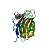


 Mass: 62.068 Da / Num. of mol.: 1 / Source method: obtained synthetically / Formula: C2H6O2
 Mass: 22.990 Da / Num. of mol.: 1 / Source method: obtained synthetically / Formula: Na
 Mass: 134.087 Da / Num. of mol.: 1 / Source method: obtained synthetically / Formula: C4H6O5
 Mass: 18.015 Da / Num. of mol.: 219 / Source method: isolated from a natural source / Formula: H2O
 Sample preparation
 Processing