| Entry | Database: PDB / ID: 7t7t
|
|---|
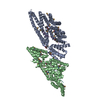
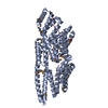
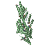
| Title | Structure of TSK/BRU1 bound to histone H3.1 |
|---|
 Components Components | - Histone H3.1
- Protein TONSOKU
|
|---|
 Keywords Keywords | NUCLEAR PROTEIN / Epigenetic protein / H3.1 reader / nucleosome |
|---|
| Function / homology |  Function and homology information Function and homology information
meristem structural organization / response to DNA damage checkpoint signaling / chromocenter / plastid / epigenetic regulation of gene expression / structural constituent of chromatin / nucleosome / chromosome / histone binding / protein heterodimerization activity ...meristem structural organization / response to DNA damage checkpoint signaling / chromocenter / plastid / epigenetic regulation of gene expression / structural constituent of chromatin / nucleosome / chromosome / histone binding / protein heterodimerization activity / DNA repair / DNA binding / extracellular region / nucleoplasm / nucleus / plasma membrane / cytosolSimilarity search - Function Protein TONSOKU / Leucine rich repeat, ribonuclease inhibitor type / Tetratricopeptide repeat / Tetratricopeptide repeat / TPR repeat region circular profile. / TPR repeat profile. / Tetratricopeptide repeats / Tetratricopeptide repeat / Leucine-rich repeat domain superfamily / Histone H3 signature 1. ...Protein TONSOKU / Leucine rich repeat, ribonuclease inhibitor type / Tetratricopeptide repeat / Tetratricopeptide repeat / TPR repeat region circular profile. / TPR repeat profile. / Tetratricopeptide repeats / Tetratricopeptide repeat / Leucine-rich repeat domain superfamily / Histone H3 signature 1. / Histone H3 signature 2. / Histone H3 / Histone H3/CENP-A / Histone H2A/H2B/H3 / Core histone H2A/H2B/H3/H4 domain / Histone-fold / Tetratricopeptide-like helical domain superfamilySimilarity search - Domain/homology |
|---|
| Biological species |   Citrus unshiu (satsuma mandarin) Citrus unshiu (satsuma mandarin)
 Arabidopsis thaliana (thale cress) Arabidopsis thaliana (thale cress) |
|---|
| Method |  X-RAY DIFFRACTION / SYNCHROTRON / SAD / Resolution: 3.17 Å X-RAY DIFFRACTION / SYNCHROTRON / SAD / Resolution: 3.17 Å |
|---|
 Authors Authors | Davarinejad, H. / Couture, J.F. |
|---|
| Funding support |  Canada, 1items Canada, 1items | Organization | Grant number | Country |
|---|
| Natural Sciences and Engineering Research Council (NSERC, Canada) | | Canada |
|
|---|
 Citation Citation | Journal: Science / Year: 2022
Title: The histone H3.1 variant regulates TONSOKU-mediated DNA repair during replication.
Authors: Davarinejad, H. / Huang, Y.C. / Mermaz, B. / LeBlanc, C. / Poulet, A. / Thomson, G. / Joly, V. / Munoz, M. / Arvanitis-Vigneault, A. / Valsakumar, D. / Villarino, G. / Ross, A. / Rotstein, B. ...Authors: Davarinejad, H. / Huang, Y.C. / Mermaz, B. / LeBlanc, C. / Poulet, A. / Thomson, G. / Joly, V. / Munoz, M. / Arvanitis-Vigneault, A. / Valsakumar, D. / Villarino, G. / Ross, A. / Rotstein, B.H. / Alarcon, E.I. / Brunzelle, J.S. / Voigt, P. / Dong, J. / Couture, J.F. / Jacob, Y. |
|---|
| History | | Deposition | Dec 15, 2021 | Deposition site: RCSB / Processing site: RCSB |
|---|
| Revision 1.0 | Mar 30, 2022 | Provider: repository / Type: Initial release |
|---|
| Revision 1.1 | Nov 6, 2024 | Group: Data collection / Structure summary
Category: chem_comp_atom / chem_comp_bond ...chem_comp_atom / chem_comp_bond / pdbx_entry_details / pdbx_modification_feature
Item: _pdbx_entry_details.has_protein_modification |
|---|
|
|---|
 Movie
Movie Controller


 Open data
 Basic information
 Structure visualization
 Downloads & links
 Other downloads
 PDBj
PDBj


 Assembly