 Movie
Movie Controller

[English] 日本語
 Yorodumi
- PDB-7t7h: Crystal structure of Vaccinia Virus decapping enzyme D9 in comple... -
+ Open data

- Basic information
| Entry | Database: PDB / ID: 7t7h | ||||||
|---|---|---|---|---|---|---|---|
| Title | Crystal structure of Vaccinia Virus decapping enzyme D9 in complex with inhibitor CP100356 | ||||||
 Components Components | DNA repair NTP-phosphohydrolase | ||||||
 Keywords Keywords | VIRAL PROTEIN/VIRAL PROTEIN INHIBITOR / decapping enzyme / nudix / inhibitor / VIRAL PROTEIN-VIRAL PROTEIN INHIBITOR complex | ||||||
| Function / homology | Viral protein D9 / NUDIX domain / Nudix hydrolase domain profile. / NUDIX hydrolase domain / NUDIX hydrolase-like domain superfamily / hydrolase activity / Chem-G7R / NTP-phosphohydrolase Function and homology information Function and homology information | ||||||
| Biological species |  Vaccinia virus Western Reserve Vaccinia virus Western Reserve | ||||||
| Method |  X-RAY DIFFRACTION / SYNCHROTRON / MOLECULAR REPLACEMENT / Resolution: 1.78000262178 Å X-RAY DIFFRACTION / SYNCHROTRON / MOLECULAR REPLACEMENT / Resolution: 1.78000262178 Å | ||||||
 Authors Authors | Peters, J.K. / Gross, J.D. | ||||||
| Funding support |  United States, 1items United States, 1items
| ||||||
 Citation Citation | Journal: Acs Chem.Biol. / Year: 2022 Title: Fluorescence-Based Activity Screening Assay Reveals Small Molecule Inhibitors of Vaccinia Virus mRNA Decapping Enzyme D9. Authors: Bednarczyk, M. / Peters, J.K. / Kasprzyk, R. / Starek, J. / Warminski, M. / Spiewla, T. / Mugridge, J.S. / Gross, J.D. / Jemielity, J. / Kowalska, J. | ||||||
| History |
|
- Structure visualization
| Structure viewer | Molecule: MolmilJmol/JSmol |
|---|
- Downloads & links
-Download
| PDBx/mmCIF format | 7t7h.cif.gz | 229.4 KB | Display | PDBx/mmCIF format |
|---|---|---|---|---|
| PDB format | pdb7t7h.ent.gz | 151.9 KB | Display | PDB format |
| PDBx/mmJSON format | 7t7h.json.gz | Tree view | PDBx/mmJSON format | |
| Others |  Other downloads Other downloads |
-Validation report
| Summary document | 7t7h_validation.pdf.gz | 1.1 MB | Display | wwPDB validaton report |
|---|---|---|---|---|
| Full document | 7t7h_full_validation.pdf.gz | 1.1 MB | Display | |
| Data in XML | 7t7h_validation.xml.gz | 21.8 KB | Display | |
| Data in CIF | 7t7h_validation.cif.gz | 29.3 KB | Display | |
| Arichive directory | https://data.pdbj.org/pub/pdb/validation_reports/t7/7t7hftp://data.pdbj.org/pub/pdb/validation_reports/t7/7t7h | HTTPS FTP |
-Related structure data
| Related structure data | 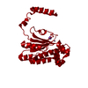 7sevS S: Starting model for refinement |
|---|---|
| Similar structure data |
-Links
 PDBj
PDBj

- Assembly
| Deposited unit | 
| |||||||||||||||||||||||||||||||||||||||||||||||||||||||||||||||||||||||||||||||||||
|---|---|---|---|---|---|---|---|---|---|---|---|---|---|---|---|---|---|---|---|---|---|---|---|---|---|---|---|---|---|---|---|---|---|---|---|---|---|---|---|---|---|---|---|---|---|---|---|---|---|---|---|---|---|---|---|---|---|---|---|---|---|---|---|---|---|---|---|---|---|---|---|---|---|---|---|---|---|---|---|---|---|---|---|---|
| 1 | 
| |||||||||||||||||||||||||||||||||||||||||||||||||||||||||||||||||||||||||||||||||||
| 2 | 
| |||||||||||||||||||||||||||||||||||||||||||||||||||||||||||||||||||||||||||||||||||
| Unit cell |
| |||||||||||||||||||||||||||||||||||||||||||||||||||||||||||||||||||||||||||||||||||
| Noncrystallographic symmetry (NCS) | NCS domain:
NCS domain segments: Ens-ID: 1
|
-Components
| #1: Protein | Mass: 26102.078 Da / Num. of mol.: 2 Source method: isolated from a genetically manipulated source Source: (gene. exp.) Vaccinia virus Western ReserveGene: D9R, VACV_BRZ_SERRO2_112, VACV_CTGV_ALEH2_114, VACV_CTGV_CG04_114, VACV_CTGV_MI233-114, VACV_CTGV_VI04_114, VAC_IHDW1_116, VAC_TKT3_104, VAC_TKT4_104, VAC_TP3_119, VAC_TP5_119, VACV_CTGV_CM01_ ...Gene: D9R, VACV_BRZ_SERRO2_112, VACV_CTGV_ALEH2_114, VACV_CTGV_CG04_114, VACV_CTGV_MI233-114, VACV_CTGV_VI04_114, VAC_IHDW1_116, VAC_TKT3_104, VAC_TKT4_104, VAC_TP3_119, VAC_TP5_119, VACV_CTGV_CM01_114, VACV_TT10_141, VACV_TT11_141, VACV_TT12_141, VACV_TT8_141, VACV_TT9_141 Production host:  #2: Chemical |   Mass: 22.990 Da / Num. of mol.: 2 / Source method: obtained synthetically / Formula: Na Mass: 22.990 Da / Num. of mol.: 2 / Source method: obtained synthetically / Formula: Na#3: Chemical |   Mass: 560.641 Da / Num. of mol.: 2 / Source method: isolated from a natural source / Formula: C31H36N4O6 / Feature type: SUBJECT OF INVESTIGATION Mass: 560.641 Da / Num. of mol.: 2 / Source method: isolated from a natural source / Formula: C31H36N4O6 / Feature type: SUBJECT OF INVESTIGATION#4: Water | ChemComp-HOH / |  Mass: 18.015 Da / Num. of mol.: 194 / Source method: isolated from a natural source / Formula: H2O Mass: 18.015 Da / Num. of mol.: 194 / Source method: isolated from a natural source / Formula: H2OHas ligand of interest | Y | |
|---|
-Experimental details
-Experiment
| Experiment | Method: X-RAY DIFFRACTION / Number of used crystals: 1 |
|---|
- Sample preparation
| Crystal | Density Matthews: 2.17 Å3/Da / Density % sol: 43.24 % |
|---|---|
| Crystal grow | Temperature: 298 K / Method: vapor diffusion, hanging drop / Details: 20% PEG 3350, 100mM NaF |
-Data collection
| Diffraction | Mean temperature: 100 K / Serial crystal experiment: N |
|---|---|
| Diffraction source | Source: SYNCHROTRON / Site: ALS / Beamline: 8.3.1 / Wavelength: 1.11583 Å |
| Detector | Type: DECTRIS PILATUS3 S 6M / Detector: PIXEL / Date: Oct 15, 2020 |
| Radiation | Protocol: SINGLE WAVELENGTH / Monochromatic (M) / Laue (L): M / Scattering type: x-ray |
| Radiation wavelength | Wavelength: 1.11583 Å / Relative weight: 1 |
| Reflection | Resolution: 1.78→62.7732227509 Å / Num. obs: 44130 / % possible obs: 99.79 % / Redundancy: 12.9 % / Biso Wilson estimate: 33.2653021128 Å2 / Rmerge(I) obs: 0.04373 / Rrim(I) all: 0.04555 / Net I/σ(I): 30.35 |
| Reflection shell | Resolution: 1.78→1.844 Å / Rmerge(I) obs: 0.9509 / Num. unique obs: 4244 |
- Processing
| Software | Name: PHENIX / Version: 1.9_1692 / Classification: refinement | |||||||||||||||||||||||||||||||||||||||||||||||||||||||||||||||||||||||||||||||||||||||||||||||||||||||||
|---|---|---|---|---|---|---|---|---|---|---|---|---|---|---|---|---|---|---|---|---|---|---|---|---|---|---|---|---|---|---|---|---|---|---|---|---|---|---|---|---|---|---|---|---|---|---|---|---|---|---|---|---|---|---|---|---|---|---|---|---|---|---|---|---|---|---|---|---|---|---|---|---|---|---|---|---|---|---|---|---|---|---|---|---|---|---|---|---|---|---|---|---|---|---|---|---|---|---|---|---|---|---|---|---|---|---|
| Refinement | Method to determine structure: MOLECULAR REPLACEMENT Starting model: 7SEV Resolution: 1.78000262178→62.7732227509 Å / SU ML: 0.176396858988 / Cross valid method: FREE R-VALUE / σ(F): 1.35113385529 / Phase error: 21.7555778038
| |||||||||||||||||||||||||||||||||||||||||||||||||||||||||||||||||||||||||||||||||||||||||||||||||||||||||
| Solvent computation | Shrinkage radii: 0.9 Å / VDW probe radii: 1.11 Å / Solvent model: FLAT BULK SOLVENT MODEL | |||||||||||||||||||||||||||||||||||||||||||||||||||||||||||||||||||||||||||||||||||||||||||||||||||||||||
| Displacement parameters | Biso mean: 49.0041316931 Å2 | |||||||||||||||||||||||||||||||||||||||||||||||||||||||||||||||||||||||||||||||||||||||||||||||||||||||||
| Refinement step | Cycle: LAST / Resolution: 1.78000262178→62.7732227509 Å
| |||||||||||||||||||||||||||||||||||||||||||||||||||||||||||||||||||||||||||||||||||||||||||||||||||||||||
| Refine LS restraints |
| |||||||||||||||||||||||||||||||||||||||||||||||||||||||||||||||||||||||||||||||||||||||||||||||||||||||||
| LS refinement shell |
| |||||||||||||||||||||||||||||||||||||||||||||||||||||||||||||||||||||||||||||||||||||||||||||||||||||||||
| Refinement TLS params. | Method: refined / Origin x: -23.5363735217 Å / Origin y: 11.1709680487 Å / Origin z: -14.3823393687 Å
| |||||||||||||||||||||||||||||||||||||||||||||||||||||||||||||||||||||||||||||||||||||||||||||||||||||||||
| Refinement TLS group | Selection details: all |
