 Movie
Movie Controller

[English] 日本語
 Yorodumi
- PDB-7t68: Co-crystal structure of Chaetomium glucosidase with compound UV-5 -
+ Open data

- Basic information
| Entry | Database: PDB / ID: 7t68 | ||||||
|---|---|---|---|---|---|---|---|
| Title | Co-crystal structure of Chaetomium glucosidase with compound UV-5 | ||||||
 Components Components | Chaetomium alpha glucosidase | ||||||
 Keywords Keywords | HYDROLASE/INHIBITOR / alpha glucosidase I / Hydrolase / Inhibitor complex / HYDROLASE-INHIBITOR complex | ||||||
| Function / homology |  Function and homology information Function and homology informationmannosyl-oligosaccharide glucosidase / Glc3Man9GlcNAc2 oligosaccharide glucosidase activity / oligosaccharide metabolic process / protein N-linked glycosylation / endoplasmic reticulum membrane Similarity search - Function | ||||||
| Biological species |  Chaetomium thermophilum var. thermophilum DSM 1495 (fungus) Chaetomium thermophilum var. thermophilum DSM 1495 (fungus) | ||||||
| Method |  X-RAY DIFFRACTION / SYNCHROTRON / MOLECULAR REPLACEMENT / Resolution: 2.32 Å X-RAY DIFFRACTION / SYNCHROTRON / MOLECULAR REPLACEMENT / Resolution: 2.32 Å | ||||||
 Authors Authors | Karade, S.S. / Mariuzza, R.A. | ||||||
| Funding support | 1items
| ||||||
 Citation Citation | Journal: Biochemistry / Year: 2022 Title: Identification of Endoplasmic Reticulum alpha-Glucosidase I from a Thermophilic Fungus as a Platform for Structure-Guided Antiviral Drug Design. Authors: Karade, S.S. / Kolesnikov, A. / Treston, A.M. / Mariuzza, R.A. | ||||||
| History |
|
- Structure visualization
| Structure viewer | Molecule: MolmilJmol/JSmol |
|---|
- Downloads & links
-Download
| PDBx/mmCIF format | 7t68.cif.gz | 324.7 KB | Display | PDBx/mmCIF format |
|---|---|---|---|---|
| PDB format | pdb7t68.ent.gz | Display | PDB format | |
| PDBx/mmJSON format | 7t68.json.gz | Tree view | PDBx/mmJSON format | |
| Others |  Other downloads Other downloads |
-Validation report
| Summary document | 7t68_validation.pdf.gz | 983.1 KB | Display | wwPDB validaton report |
|---|---|---|---|---|
| Full document | 7t68_full_validation.pdf.gz | 1006.3 KB | Display | |
| Data in XML | 7t68_validation.xml.gz | 57.3 KB | Display | |
| Data in CIF | 7t68_validation.cif.gz | 79.7 KB | Display | |
| Arichive directory | https://data.pdbj.org/pub/pdb/validation_reports/t6/7t68ftp://data.pdbj.org/pub/pdb/validation_reports/t6/7t68 | HTTPS FTP |
-Related structure data
| Related structure data | 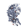 7t66C  7t6wC  7t8vC 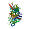 4j5tS S: Starting model for refinement C: citing same article ( |
|---|---|
| Similar structure data |
-Links
 PDBj
PDBj
- Assembly
| Deposited unit | 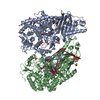
| ||||||||
|---|---|---|---|---|---|---|---|---|---|
| 1 | 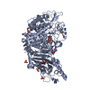
| ||||||||
| 2 | 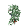
| ||||||||
| Unit cell |
|
-Components
-Protein / Sugars , 2 types, 3 molecules AB
| #1: Protein | Mass: 93319.812 Da / Num. of mol.: 2 Source method: isolated from a genetically manipulated source Source: (gene. exp.) Chaetomium thermophilum var. thermophilum DSM 1495 (fungus)Strain: DSM 1495 / CBS 144.50 / IMI 039719 / Gene: CTHT_0061620 / Cell line (production host): Expi-HEK293 / Production host:  Homo sapiens (human) / References: UniProt: G0SFD1 Homo sapiens (human) / References: UniProt: G0SFD1#5: Sugar | ChemComp-NAG / |  Type: D-saccharide, beta linking / Mass: 221.208 Da / Num. of mol.: 1 Type: D-saccharide, beta linking / Mass: 221.208 Da / Num. of mol.: 1Source method: isolated from a genetically manipulated source Formula: C8H15NO6 |
|---|
-Non-polymers , 5 types, 313 molecules 


| #2: Chemical |  Mass: 424.452 Da / Num. of mol.: 2 / Source method: obtained synthetically / Formula: C18H28N6O6 / Feature type: SUBJECT OF INVESTIGATION Mass: 424.452 Da / Num. of mol.: 2 / Source method: obtained synthetically / Formula: C18H28N6O6 / Feature type: SUBJECT OF INVESTIGATION#3: Chemical |  Mass: 92.094 Da / Num. of mol.: 2 / Source method: obtained synthetically / Formula: C3H8O3 Mass: 92.094 Da / Num. of mol.: 2 / Source method: obtained synthetically / Formula: C3H8O3#4: Chemical | ChemComp-SO4 /  Mass: 96.063 Da / Num. of mol.: 13 / Source method: obtained synthetically / Formula: SO4 Mass: 96.063 Da / Num. of mol.: 13 / Source method: obtained synthetically / Formula: SO4#6: Chemical | ChemComp-BTB / |  Mass: 209.240 Da / Num. of mol.: 1 / Source method: obtained synthetically / Formula: C8H19NO5 / Comment: pH buffer*YM Mass: 209.240 Da / Num. of mol.: 1 / Source method: obtained synthetically / Formula: C8H19NO5 / Comment: pH buffer*YM#7: Water | ChemComp-HOH / | Mass: 18.015 Da / Num. of mol.: 295 / Source method: isolated from a natural source / Formula: H2O |
|---|
-Details
| Has ligand of interest | Y |
|---|---|
| Has protein modification | Y |
-Experimental details
-Experiment
| Experiment | Method: X-RAY DIFFRACTION / Number of used crystals: 1 |
|---|
- Sample preparation
| Crystal | Density Matthews: 2.91 Å3/Da / Density % sol: 57.76 % |
|---|---|
| Crystal grow | Temperature: 297 K / Method: vapor diffusion, hanging drop / pH: 6.5 / Details: 0.1 Bis Tris pH 6.5, 1.6-2.0 M Ammonium sulfate |
-Data collection
| Diffraction | Mean temperature: 100 K / Serial crystal experiment: N |
|---|---|
| Diffraction source | Source: SYNCHROTRON / Site: SSRL  / Beamline: BL9-2 / Wavelength: 0.9794 Å / Beamline: BL9-2 / Wavelength: 0.9794 Å |
| Detector | Type: DECTRIS PILATUS 6M / Detector: PIXEL / Date: Jun 11, 2021 |
| Radiation | Monochromator: Si(111) and Si(220) double crystal / Protocol: SINGLE WAVELENGTH / Monochromatic (M) / Laue (L): M / Scattering type: x-ray |
| Radiation wavelength | Wavelength: 0.9794 Å / Relative weight: 1 |
| Reflection | Resolution: 2.32→46.72 Å / Num. obs: 93750 / % possible obs: 99.68 % / Redundancy: 6.7 % / CC1/2: 0.998 / CC star: 1 / Rmerge(I) obs: 0.08142 / Rpim(I) all: 0.03383 / Rrim(I) all: 0.08831 / Net I/σ(I): 9.63 |
| Reflection shell | Resolution: 2.32→2.404 Å / Redundancy: 6.3 % / Rmerge(I) obs: 0.6095 / Mean I/σ(I) obs: 1.85 / Num. unique obs: 9178 / CC1/2: 0.842 / CC star: 0.956 / Rpim(I) all: 0.2616 / Rrim(I) all: 0.6646 / % possible all: 99.01 |
- Processing
| Software |
| |||||||||||||||||||||||||||||||||||||||||||||
|---|---|---|---|---|---|---|---|---|---|---|---|---|---|---|---|---|---|---|---|---|---|---|---|---|---|---|---|---|---|---|---|---|---|---|---|---|---|---|---|---|---|---|---|---|---|---|
| Refinement | Method to determine structure: MOLECULAR REPLACEMENT Starting model: 4J5T Resolution: 2.32→46.72 Å / Cor.coef. Fo:Fc: 0.955 / Cor.coef. Fo:Fc free: 0.936 / SU B: 6.83 / SU ML: 0.16 / Cross valid method: THROUGHOUT / σ(F): 0 / ESU R: 0.258 / ESU R Free: 0.208 / Stereochemistry target values: MAXIMUM LIKELIHOOD / Details: U VALUES : REFINED INDIVIDUALLY
| |||||||||||||||||||||||||||||||||||||||||||||
| Solvent computation | Ion probe radii: 0.8 Å / Shrinkage radii: 0.8 Å / VDW probe radii: 1.2 Å / Solvent model: MASK | |||||||||||||||||||||||||||||||||||||||||||||
| Displacement parameters | Biso max: 122.59 Å2 / Biso mean: 40.862 Å2 / Biso min: 19.44 Å2
| |||||||||||||||||||||||||||||||||||||||||||||
| Refinement step | Cycle: final / Resolution: 2.32→46.72 Å
| |||||||||||||||||||||||||||||||||||||||||||||
| Refine LS restraints |
| |||||||||||||||||||||||||||||||||||||||||||||
| LS refinement shell | Resolution: 2.321→2.381 Å / Rfactor Rfree error: 0 / Total num. of bins used: 20
|
