Movie
Movie Controller

[English] 日本語
 Yorodumi
- PDB-7t5q: Cryo-EM Structure of a Transition State of Arp2/3 Complex Activation -
+ Open data

- Basic information
| Entry | Database: PDB / ID: 7t5q | ||||||||||||
|---|---|---|---|---|---|---|---|---|---|---|---|---|---|
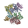
| Title | Cryo-EM Structure of a Transition State of Arp2/3 Complex Activation | ||||||||||||
 Components Components |
| ||||||||||||
 Keywords Keywords | CONTRACTILE PROTEIN / actin / ATPase / actin related protein / arp / cytoskeleton / Arp2-3 complex / actin nucleation / actin branching / CapZ | ||||||||||||
| Function / homology |  Function and homology information Function and homology informationnegative regulation of membrane tubulation / spindle localization / membrane invagination / plasma membrane tubulation / muscle cell projection membrane / EPHB-mediated forward signaling / Regulation of actin dynamics for phagocytic cup formation / RHO GTPases Activate WASPs and WAVEs / postsynaptic actin cytoskeleton organization / Arp2/3 protein complex ...negative regulation of membrane tubulation / spindle localization / membrane invagination / plasma membrane tubulation / muscle cell projection membrane / EPHB-mediated forward signaling / Regulation of actin dynamics for phagocytic cup formation / RHO GTPases Activate WASPs and WAVEs / postsynaptic actin cytoskeleton organization / Arp2/3 protein complex / negative regulation of lymphocyte migration / actin nucleation / positive regulation of clathrin-dependent endocytosis / Arp2/3 complex-mediated actin nucleation / vesicle transport along actin filament / F-actin capping protein complex / WASH complex / regulation of cell projection assembly / actin cap / postsynapse organization / vesicle organization / regulation of actin filament polymerization / Clathrin-mediated endocytosis / vesicle budding from membrane / cell junction assembly / positive regulation of chemotaxis / dendritic spine morphogenesis / barbed-end actin filament capping / actin polymerization or depolymerization / RHOD GTPase cycle / regulation of cell morphogenesis / RHOF GTPase cycle / COPI-independent Golgi-to-ER retrograde traffic / regulation of postsynapse organization / Neutrophil degranulation / positive regulation of filopodium assembly / protein-containing complex localization / lamellipodium assembly / Sensory processing of sound by inner hair cells of the cochlea / cytoskeletal motor activator activity / myosin heavy chain binding / tropomyosin binding / actin filament bundle / troponin I binding / filamentous actin / mesenchyme migration / positive regulation of actin filament polymerization / cell leading edge / cortical cytoskeleton / skeletal muscle myofibril / brush border / actin filament bundle assembly / Advanced glycosylation endproduct receptor signaling / striated muscle thin filament / skeletal muscle thin filament assembly / cilium assembly / actin monomer binding / sperm head-tail coupling apparatus / positive regulation of double-strand break repair via homologous recombination / COPI-mediated anterograde transport / positive regulation of lamellipodium assembly / skeletal muscle fiber development / cytoskeletal protein binding / cell projection / stress fiber / titin binding / actin filament polymerization / positive regulation of substrate adhesion-dependent cell spreading / cytoskeleton organization / MHC class II antigen presentation / Gene and protein expression by JAK-STAT signaling after Interleukin-12 stimulation / HSP90 chaperone cycle for steroid hormone receptors (SHR) in the presence of ligand / hippocampal mossy fiber to CA3 synapse / sarcomere / response to bacterium / filopodium / actin filament / structural constituent of cytoskeleton / Hydrolases; Acting on acid anhydrides; Acting on acid anhydrides to facilitate cellular and subcellular movement / Schaffer collateral - CA1 synapse / calcium-dependent protein binding / actin filament binding / cell migration / regulation of protein localization / lamellipodium / actin cytoskeleton / synaptic vesicle membrane / site of double-strand break / Factors involved in megakaryocyte development and platelet production / actin binding / actin cytoskeleton organization / cell body / protein-containing complex assembly / cytoplasmic vesicle / cell cortex / cytoskeleton / protein-macromolecule adaptor activity / endosome / neuron projection / postsynaptic density Similarity search - Function | ||||||||||||
| Biological species |  Homo sapiens (human) Homo sapiens (human) | ||||||||||||
| Method | ELECTRON MICROSCOPY / single particle reconstruction / cryo EM / Resolution: 3.4 Å | ||||||||||||
 Authors Authors | Rebowski, G. / van Eeuwen, T. / Boczkowska, M. / Dominguez, R. | ||||||||||||
| Funding support |  United States, 3items United States, 3items
| ||||||||||||
 Citation Citation | Journal: Proc Natl Acad Sci U S A / Year: 2023 Title: Transition State of Arp2/3 Complex Activation by Actin-Bound Dimeric Nucleation-Promoting Factor. Authors: Trevor van Eeuwen / Malgorzata Boczkowska / Grzegorz Rebowski / Peter J Carman / Fred E Fregoso / Roberto Dominguez / Abstract: Arp2/3 complex generates branched actin networks that drive fundamental processes such as cell motility and cytokinesis. The complex comprises seven proteins, including actin-related proteins (Arps) ...Arp2/3 complex generates branched actin networks that drive fundamental processes such as cell motility and cytokinesis. The complex comprises seven proteins, including actin-related proteins (Arps) 2 and 3 and five scaffolding proteins (ArpC1-ArpC5) that mediate interactions with a pre-existing (mother) actin filament at the branch junction. Arp2/3 complex exists in two main conformations, inactive with the Arps interacting end-to-end and active with the Arps interacting side-by-side like subunits of the short-pitch helix of the actin filament. Several cofactors drive the transition toward the active state, including ATP binding to the Arps, WASP-family nucleation-promoting factors (NPFs), actin monomers, and binding of Arp2/3 complex to the mother filament. The precise contribution of each cofactor to activation is poorly understood. We report the 3.32-Å resolution cryo-electron microscopy structure of a transition state of Arp2/3 complex activation with bound constitutively dimeric NPF. Arp2/3 complex-binding region of the NPF N-WASP was fused C-terminally to the α and β subunits of the CapZ heterodimer. One arm of the NPF dimer binds Arp2 and the other binds actin and Arp3. The conformation of the complex is intermediate between those of inactive and active Arp2/3 complex. Arp2, Arp3, and actin also adopt intermediate conformations between monomeric (G-actin) and filamentous (F-actin) states, but only actin hydrolyzes ATP. In solution, the transition complex is kinetically shifted toward the short-pitch conformation and has higher affinity for F-actin than inactive Arp2/3 complex. The results reveal how all the activating cofactors contribute in a coordinated manner toward Arp2/3 complex activation. | ||||||||||||
| History |
|
- Structure visualization
| Structure viewer | Molecule: MolmilJmol/JSmol |
|---|
- Downloads & links
-Download
| PDBx/mmCIF format | 7t5q.cif.gz | 554.9 KB | Display | PDBx/mmCIF format |
|---|---|---|---|---|
| PDB format | pdb7t5q.ent.gz | 438.7 KB | Display | PDB format |
| PDBx/mmJSON format | 7t5q.json.gz | Tree view | PDBx/mmJSON format | |
| Others |  Other downloads Other downloads |
-Validation report
| Arichive directory | https://data.pdbj.org/pub/pdb/validation_reports/t5/7t5qftp://data.pdbj.org/pub/pdb/validation_reports/t5/7t5q | HTTPS FTP |
|---|
-Related structure data
| Related structure data |  25707MC M: map data used to model this data C: citing same article ( |
|---|---|
| Similar structure data |
-Links
 PDBj
PDBj


- Assembly
| Deposited unit | 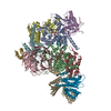
|
|---|---|
| 1 |
|
-Components
-Actin-related protein ... , 7 types, 7 molecules ABCDEFG
| #1: Protein | Mass: 47428.031 Da / Num. of mol.: 1 / Source method: isolated from a natural source / Source: (natural) |
|---|---|
| #2: Protein | Mass: 44818.711 Da / Num. of mol.: 1 / Source method: isolated from a natural source / Source: (natural) |
| #3: Protein | Mass: 41594.238 Da / Num. of mol.: 1 / Source method: isolated from a natural source / Source: (natural) |
| #4: Protein | Mass: 34402.043 Da / Num. of mol.: 1 / Source method: isolated from a natural source / Source: (natural) |
| #5: Protein | Mass: 20572.666 Da / Num. of mol.: 1 / Source method: isolated from a natural source / Source: (natural) |
| #6: Protein | Mass: 19697.047 Da / Num. of mol.: 1 / Source method: isolated from a natural source / Source: (natural) |
| #7: Protein | Mass: 16251.308 Da / Num. of mol.: 1 / Source method: isolated from a natural source / Source: (natural) |
-Protein , 1 types, 1 molecules H
| #8: Protein | Mass: 41875.633 Da / Num. of mol.: 1 / Source method: isolated from a natural source / Source: (natural) |
|---|
-F-actin-capping protein subunit ... , 2 types, 2 molecules IJ
| #9: Protein | Mass: 39600.566 Da / Num. of mol.: 1 Source method: isolated from a genetically manipulated source Source: (gene. exp.) Homo sapiens (human), (gene. exp.) Gene: CAPZA1, Wasl / Production host:  |
|---|---|
| #10: Protein | Mass: 37470.754 Da / Num. of mol.: 1 Source method: isolated from a genetically manipulated source Source: (gene. exp.) Homo sapiens (human), (gene. exp.) Gene: CAPZB, Wasl / Production host: |
-Non-polymers , 3 types, 6 molecules 


| #11: Chemical |  Mass: 24.305 Da / Num. of mol.: 3 / Source method: obtained synthetically / Formula: Mg Mass: 24.305 Da / Num. of mol.: 3 / Source method: obtained synthetically / Formula: Mg#12: Chemical |  Mass: 507.181 Da / Num. of mol.: 2 / Source method: obtained synthetically / Formula: C10H16N5O13P3 / Comment: ATP, energy-carrying molecule*YM Mass: 507.181 Da / Num. of mol.: 2 / Source method: obtained synthetically / Formula: C10H16N5O13P3 / Comment: ATP, energy-carrying molecule*YM#13: Chemical | ChemComp-ADP / |  Mass: 427.201 Da / Num. of mol.: 1 / Source method: obtained synthetically / Formula: C10H15N5O10P2 / Comment: ADP, energy-carrying molecule*YM Mass: 427.201 Da / Num. of mol.: 1 / Source method: obtained synthetically / Formula: C10H15N5O10P2 / Comment: ADP, energy-carrying molecule*YM |
|---|
-Details
| Has ligand of interest | N |
|---|---|
| Has protein modification | Y |
-Experimental details
-Experiment
| Experiment | Method: ELECTRON MICROSCOPY |
|---|---|
| EM experiment | Aggregation state: PARTICLE / 3D reconstruction method: single particle reconstruction |
- Sample preparation
| Component |
| |||||||||||||||||||||||||||||||||||
|---|---|---|---|---|---|---|---|---|---|---|---|---|---|---|---|---|---|---|---|---|---|---|---|---|---|---|---|---|---|---|---|---|---|---|---|---|
| Molecular weight | Value: 0.2489 MDa / Experimental value: NO | |||||||||||||||||||||||||||||||||||
| Source (natural) |
| |||||||||||||||||||||||||||||||||||
| Source (recombinant) |
| |||||||||||||||||||||||||||||||||||
| Buffer solution | pH: 7 | |||||||||||||||||||||||||||||||||||
| Buffer component |
| |||||||||||||||||||||||||||||||||||
| Specimen | Conc.: 5 mg/ml / Embedding applied: NO / Shadowing applied: NO / Staining applied: NO / Vitrification applied: YES / Details: This sample was monodisperse. | |||||||||||||||||||||||||||||||||||
| Vitrification | Instrument: LEICA EM CPC / Cryogen name: ETHANE Details: Grids were manually blotted for 3 seconds with Whatman 41 filter paper and manually plunged using a Leica EM CPC manual plunger. |
- Electron microscopy imaging
| Experimental equipment |  Model: Titan Krios / Image courtesy: FEI Company |
|---|---|
| Microscopy | Model: FEI TITAN KRIOS Details: Data were collected in super-resolution mode with an illuminated area of 1.01 um, nominal dose of 40 e-/A^2, a dose rate of 4.87 e-/s/pixel, and 2 or 5 exposures per hole by image shift. |
| Electron gun | Electron source:  FIELD EMISSION GUN / Accelerating voltage: 300 kV / Illumination mode: SPOT SCAN FIELD EMISSION GUN / Accelerating voltage: 300 kV / Illumination mode: SPOT SCAN |
| Electron lens | Mode: BRIGHT FIELD / Nominal magnification: 165000 X / Nominal defocus max: 3500 nm / Nominal defocus min: 1500 nm / Cs: 2.7 mm / C2 aperture diameter: 100 µm / Alignment procedure: COMA FREE |
| Specimen holder | Cryogen: NITROGEN / Specimen holder model: FEI TITAN KRIOS AUTOGRID HOLDER |
| Image recording | Average exposure time: 2.6 sec. / Electron dose: 50 e/Å2 / Detector mode: SUPER-RESOLUTION / Film or detector model: GATAN K3 (6k x 4k) / Num. of grids imaged: 1 / Num. of real images: 9661 |
| EM imaging optics | Energyfilter slit width: 20 eV |
| Image scans | Width: 5760 / Height: 4092 / Movie frames/image: 40 / Used frames/image: 1-40 |
- Processing
| Software | Name: PHENIX / Version: 1.20.1_4487: / Classification: refinement | |||||||||||||||||||||||||||||||||||||||||||||||||||||||
|---|---|---|---|---|---|---|---|---|---|---|---|---|---|---|---|---|---|---|---|---|---|---|---|---|---|---|---|---|---|---|---|---|---|---|---|---|---|---|---|---|---|---|---|---|---|---|---|---|---|---|---|---|---|---|---|---|
| EM software |
| |||||||||||||||||||||||||||||||||||||||||||||||||||||||
| Image processing | Details: Super resolution mode; micrographs binned during motion correction | |||||||||||||||||||||||||||||||||||||||||||||||||||||||
| CTF correction | Details: CTF correction was done in cryoSPARC for intial 2D classification and then repeated in CTFFIND4 (Relion) for classification and final reconstruction. Type: PHASE FLIPPING AND AMPLITUDE CORRECTION | |||||||||||||||||||||||||||||||||||||||||||||||||||||||
| Particle selection | Num. of particles selected: 4485066 / Details: Particles autopicked in cryoSPARC | |||||||||||||||||||||||||||||||||||||||||||||||||||||||
| Symmetry | Point symmetry: C1 (asymmetric) | |||||||||||||||||||||||||||||||||||||||||||||||||||||||
| 3D reconstruction | Resolution: 3.4 Å / Resolution method: FSC 0.143 CUT-OFF / Num. of particles: 269760 / Algorithm: FOURIER SPACE / Details: Composite map after multibody refinement / Num. of class averages: 1 / Symmetry type: POINT | |||||||||||||||||||||||||||||||||||||||||||||||||||||||
| Atomic model building | B value: 132 / Protocol: FLEXIBLE FIT / Space: REAL / Target criteria: correlation | |||||||||||||||||||||||||||||||||||||||||||||||||||||||
| Atomic model building | PDB-ID: 4JD2 Accession code: 4JD2 / Source name: PDB / Type: experimental model | |||||||||||||||||||||||||||||||||||||||||||||||||||||||
| Refine LS restraints |
|