 Movie
Movie Controller

[English] 日本語
 Yorodumi
- PDB-7t5h: Structure of rabies virus phosphoprotein C-terminal domain, wild type -
+ Open data

- Basic information
| Entry | Database: PDB / ID: 7t5h | |||||||||
|---|---|---|---|---|---|---|---|---|---|---|
| Title | Structure of rabies virus phosphoprotein C-terminal domain, wild type | |||||||||
 Components Components | Phosphoprotein | |||||||||
 Keywords Keywords | VIRAL PROTEIN / rabies / phosphoprotein / virus / lyssavirus | |||||||||
| Function / homology |  Function and homology information Function and homology informationmicrotubule-dependent intracellular transport of viral material towards nucleus / viral transcription / symbiont-mediated suppression of host JAK-STAT cascade via inhibition of STAT2 activity / symbiont-mediated suppression of host JAK-STAT cascade via inhibition of STAT1 activity / virion component / host cell / symbiont-mediated suppression of host cytoplasmic pattern recognition receptor signaling pathway via inhibition of TBK1 activity / symbiont-mediated suppression of host toll-like receptor signaling pathway / host cell cytoplasm / symbiont-mediated suppression of host type I interferon-mediated signaling pathway ...microtubule-dependent intracellular transport of viral material towards nucleus / viral transcription / symbiont-mediated suppression of host JAK-STAT cascade via inhibition of STAT2 activity / symbiont-mediated suppression of host JAK-STAT cascade via inhibition of STAT1 activity / virion component / host cell / symbiont-mediated suppression of host cytoplasmic pattern recognition receptor signaling pathway via inhibition of TBK1 activity / symbiont-mediated suppression of host toll-like receptor signaling pathway / host cell cytoplasm / symbiont-mediated suppression of host type I interferon-mediated signaling pathway / RNA-directed RNA polymerase activity / symbiont entry into host cell / host cell nucleus Similarity search - Function | |||||||||
| Biological species |  Rabies virus Nishigahara RCEH Rabies virus Nishigahara RCEH | |||||||||
| Method |  X-RAY DIFFRACTION / SYNCHROTRON / MOLECULAR REPLACEMENT / Resolution: 1.5 Å X-RAY DIFFRACTION / SYNCHROTRON / MOLECULAR REPLACEMENT / Resolution: 1.5 Å | |||||||||
 Authors Authors | Zhan, J. / Metcalfe, R.D. / Gooley, P.R. / Griffin, M.D.W. | |||||||||
| Funding support |  Australia, 2items Australia, 2items
| |||||||||
 Citation Citation | Journal: J.Virol. / Year: 2022 Title: Molecular Basis of Functional Effects of Phosphorylation of the C-Terminal Domain of the Rabies Virus P Protein. Authors: Zhan, J. / Watts, E. / Brice, A.M. / Metcalfe, R.D. / Rozario, A.M. / Sethi, A. / Yan, F. / Bell, T.D.M. / Griffin, M.D.W. / Moseley, G.W. / Gooley, P.R. | |||||||||
| History |
|
- Structure visualization
| Structure viewer | Molecule: MolmilJmol/JSmol |
|---|
- Downloads & links
-Download
| PDBx/mmCIF format | 7t5h.cif.gz | 96.3 KB | Display | PDBx/mmCIF format |
|---|---|---|---|---|
| PDB format | pdb7t5h.ent.gz | 60 KB | Display | PDB format |
| PDBx/mmJSON format | 7t5h.json.gz | Tree view | PDBx/mmJSON format | |
| Others |  Other downloads Other downloads |
-Validation report
| Arichive directory | https://data.pdbj.org/pub/pdb/validation_reports/t5/7t5hftp://data.pdbj.org/pub/pdb/validation_reports/t5/7t5h | HTTPS FTP |
|---|
-Related structure data
| Related structure data |  7t5gC  1vyiS S: Starting model for refinement C: citing same article ( |
|---|---|
| Similar structure data |
-Links
 PDBj
PDBj- Assembly
| Deposited unit | 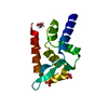
| ||||||||||||
|---|---|---|---|---|---|---|---|---|---|---|---|---|---|
| 1 |
| ||||||||||||
| Unit cell |
|
-Components
| #1: Protein | Mass: 13018.811 Da / Num. of mol.: 1 / Fragment: C-terminal domain / Mutation: C297S Source method: isolated from a genetically manipulated source Source: (gene. exp.) Rabies virus Nishigahara RCEH / Strain: Nishigahara RCEH / Production host:  | ||||||
|---|---|---|---|---|---|---|---|
| #2: Chemical | ChemComp-EDO /   Mass: 62.068 Da / Num. of mol.: 1 / Source method: obtained synthetically / Formula: C2H6O2 Mass: 62.068 Da / Num. of mol.: 1 / Source method: obtained synthetically / Formula: C2H6O2 | ||||||
| #3: Chemical |   Mass: 96.063 Da / Num. of mol.: 2 / Source method: obtained synthetically / Formula: SO4 Mass: 96.063 Da / Num. of mol.: 2 / Source method: obtained synthetically / Formula: SO4#4: Chemical | ChemComp-PO4 / |   Mass: 94.971 Da / Num. of mol.: 1 / Source method: obtained synthetically / Formula: PO4 Mass: 94.971 Da / Num. of mol.: 1 / Source method: obtained synthetically / Formula: PO4#5: Water | ChemComp-HOH / |  Mass: 18.015 Da / Num. of mol.: 103 / Source method: isolated from a natural source / Formula: H2O Mass: 18.015 Da / Num. of mol.: 103 / Source method: isolated from a natural source / Formula: H2OHas ligand of interest | N | |
-Experimental details
-Experiment
| Experiment | Method: X-RAY DIFFRACTION / Number of used crystals: 1 |
|---|
- Sample preparation
| Crystal | Density Matthews: 2.21 Å3/Da / Density % sol: 44.3 % |
|---|---|
| Crystal grow | Temperature: 281 K / Method: vapor diffusion, sitting drop / pH: 4.75 Details: 1.9 M ammonium sulfate, 0.2 M potassium sodium tartrate and 0.1 M sodium citrate (pH 4.75) |
-Data collection
| Diffraction | Mean temperature: 100 K / Serial crystal experiment: N |
|---|---|
| Diffraction source | Source: SYNCHROTRON / Site: Australian Synchrotron / Beamline: MX2 / Wavelength: 0.9537 Å |
| Detector | Type: DECTRIS EIGER X 16M / Detector: PIXEL / Date: Jun 13, 2018 |
| Radiation | Protocol: SINGLE WAVELENGTH / Monochromatic (M) / Laue (L): M / Scattering type: x-ray |
| Radiation wavelength | Wavelength: 0.9537 Å / Relative weight: 1 |
| Reflection | Resolution: 1.5→43.99 Å / Num. obs: 18982 / % possible obs: 100 % / Redundancy: 12.7 % / Biso Wilson estimate: 23 Å2 / CC1/2: 0.999 / Rmerge(I) obs: 0.085 / Rpim(I) all: 0.025 / Rrim(I) all: 0.088 / Net I/σ(I): 14.9 |
| Reflection shell | Resolution: 1.5→1.53 Å / Rmerge(I) obs: 1.463 / Mean I/σ(I) obs: 1.6 / Num. unique obs: 928 / CC1/2: 0.816 / Rpim(I) all: 0.411 / Rrim(I) all: 1.544 |
- Processing
| Software |
| ||||||||||||||||||||||||||||||||||||||||||||||||||||||||
|---|---|---|---|---|---|---|---|---|---|---|---|---|---|---|---|---|---|---|---|---|---|---|---|---|---|---|---|---|---|---|---|---|---|---|---|---|---|---|---|---|---|---|---|---|---|---|---|---|---|---|---|---|---|---|---|---|---|
| Refinement | Method to determine structure: MOLECULAR REPLACEMENT Starting model: 1vyi Resolution: 1.5→34.83 Å / SU ML: 0.1384 / Cross valid method: FREE R-VALUE / σ(F): 1.32 / Phase error: 22.9442 Stereochemistry target values: GeoStd + Monomer Library + CDL v1.2
| ||||||||||||||||||||||||||||||||||||||||||||||||||||||||
| Solvent computation | Shrinkage radii: 0.9 Å / VDW probe radii: 1.11 Å / Solvent model: FLAT BULK SOLVENT MODEL | ||||||||||||||||||||||||||||||||||||||||||||||||||||||||
| Displacement parameters | Biso mean: 32.07 Å2 | ||||||||||||||||||||||||||||||||||||||||||||||||||||||||
| Refinement step | Cycle: LAST / Resolution: 1.5→34.83 Å
| ||||||||||||||||||||||||||||||||||||||||||||||||||||||||
| Refine LS restraints |
| ||||||||||||||||||||||||||||||||||||||||||||||||||||||||
| LS refinement shell |
| ||||||||||||||||||||||||||||||||||||||||||||||||||||||||
| Refinement TLS params. | Method: refined / Origin x: 0.205659003875 Å / Origin y: -9.76543998279 Å / Origin z: 11.4348981287 Å
| ||||||||||||||||||||||||||||||||||||||||||||||||||||||||
| Refinement TLS group | Selection details: (chain 'A' and resid 190 through 297) |
