 Movie
Movie Controller


+ Open data

- Basic information
| Entry | Database: PDB / ID: 7t4x | |||||||||
|---|---|---|---|---|---|---|---|---|---|---|
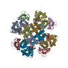
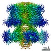
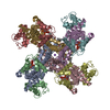
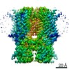
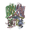
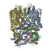
| Title | AKT1 K+ channel from A. thaliana in MSP2N2 lipid nanodisc | |||||||||
 Components Components | Potassium channel AKT1 | |||||||||
 Keywords Keywords | MEMBRANE PROTEIN / K+ channel / ion channel / hyperpolarization-activated channel / voltage-gated channel | |||||||||
| Function / homology |  Function and homology information Function and homology informationroot hair elongation / regulation of stomatal closure / response to water deprivation / inward rectifier potassium channel activity / monoatomic ion channel complex / potassium ion import across plasma membrane / response to salt stress / potassium ion transport / identical protein binding / plasma membrane Similarity search - Function | |||||||||
| Biological species |  | |||||||||
| Method | ELECTRON MICROSCOPY / single particle reconstruction / cryo EM / Resolution: 2.8 Å | |||||||||
 Authors Authors | Dickinson, M.S. / Pourmal, S. | |||||||||
| Funding support |  United States, 1items United States, 1items
| |||||||||
 Citation Citation | Journal: Biochemistry / Year: 2022 Title: Symmetry Reduction in a Hyperpolarization-Activated Homotetrameric Ion Channel. Authors: Miles Sasha Dickinson / Sergei Pourmal / Meghna Gupta / Maxine Bi / Robert M Stroud / Abstract: Plants obtain nutrients from the soil via transmembrane transporters and channels in their root hairs, from which ions radially transport in toward the xylem for distribution across the plant body. ...Plants obtain nutrients from the soil via transmembrane transporters and channels in their root hairs, from which ions radially transport in toward the xylem for distribution across the plant body. We determined structures of the hyperpolarization-activated channel AKT1 from , which mediates K uptake from the soil into plant roots. These structures of AtAKT1 embedded in lipid nanodiscs show that the channel undergoes a reduction of C4 to C2 symmetry, possibly to regulate its electrical activation. | |||||||||
| History |
|
- Structure visualization
| Movie |
Movie viewer |
|---|---|
| Structure viewer | Molecule: MolmilJmol/JSmol |
- Downloads & links
-Download
| PDBx/mmCIF format | 7t4x.cif.gz | 630.2 KB | Display | PDBx/mmCIF format |
|---|---|---|---|---|
| PDB format | pdb7t4x.ent.gz | 511.5 KB | Display | PDB format |
| PDBx/mmJSON format | 7t4x.json.gz | Tree view | PDBx/mmJSON format | |
| Others |  Other downloads Other downloads |
-Validation report
| Arichive directory | https://data.pdbj.org/pub/pdb/validation_reports/t4/7t4xftp://data.pdbj.org/pub/pdb/validation_reports/t4/7t4x | HTTPS FTP |
|---|
-Related structure data
| Related structure data |  25691MC M: map data used to model this data C: citing same article ( |
|---|---|
| Similar structure data |


-Links
 PDBj
PDBj


- Assembly
| Deposited unit | 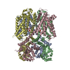
|
|---|---|
| 1 |
|
-Components
| #1: Protein | Mass: 97109.625 Da / Num. of mol.: 4 Source method: isolated from a genetically manipulated source Source: (gene. exp.)  Homo sapiens (human) / References: UniProt: Q38998 Homo sapiens (human) / References: UniProt: Q38998#2: Chemical | ChemComp-K /   Mass: 39.098 Da / Num. of mol.: 5 / Source method: obtained synthetically / Formula: K / Feature type: SUBJECT OF INVESTIGATION Mass: 39.098 Da / Num. of mol.: 5 / Source method: obtained synthetically / Formula: K / Feature type: SUBJECT OF INVESTIGATION#3: Chemical |   Mass: 734.039 Da / Num. of mol.: 2 / Source method: obtained synthetically / Formula: C40H80NO8P / Feature type: SUBJECT OF INVESTIGATION / Comment: phospholipid*YM Mass: 734.039 Da / Num. of mol.: 2 / Source method: obtained synthetically / Formula: C40H80NO8P / Feature type: SUBJECT OF INVESTIGATION / Comment: phospholipid*YM#4: Water | ChemComp-HOH / |  Mass: 18.015 Da / Num. of mol.: 13 / Source method: isolated from a natural source / Formula: H2O Mass: 18.015 Da / Num. of mol.: 13 / Source method: isolated from a natural source / Formula: H2OHas ligand of interest | Y | Has protein modification | Y | |
|---|
-Experimental details
-Experiment
| Experiment | Method: ELECTRON MICROSCOPY |
|---|---|
| EM experiment | Aggregation state: PARTICLE / 3D reconstruction method: single particle reconstruction |
- Sample preparation
| Component | Name: Homotetramer of AKT1 subunits / Type: COMPLEX / Entity ID: #1 / Source: RECOMBINANT | |||||||||||||||
|---|---|---|---|---|---|---|---|---|---|---|---|---|---|---|---|---|
| Molecular weight | Value: 0.96989 MDa / Experimental value: NO | |||||||||||||||
| Source (natural) | Organism: | |||||||||||||||
| Source (recombinant) | Organism: Homo sapiens (human) | |||||||||||||||
| Buffer solution | pH: 7.5 | |||||||||||||||
| Buffer component |
| |||||||||||||||
| Specimen | Conc.: 0.4 mg/ml / Embedding applied: NO / Shadowing applied: NO / Staining applied: NO / Vitrification applied: YES Details: Homotetramer of AKT1 subunits in MSP2N2 lipid nanodisc | |||||||||||||||
| Specimen support | Grid material: GOLD / Grid mesh size: 300 divisions/in. / Grid type: Quantifoil R1.2/1.3 | |||||||||||||||
| Vitrification | Instrument: FEI VITROBOT MARK IV / Cryogen name: ETHANE / Humidity: 100 % / Chamber temperature: 277.15 K |
- Electron microscopy imaging
| Experimental equipment |  Model: Titan Krios / Image courtesy: FEI Company |
|---|---|
| Microscopy | Model: TFS KRIOS |
| Electron gun | Electron source:  FIELD EMISSION GUN / Accelerating voltage: 300 kV / Illumination mode: FLOOD BEAM FIELD EMISSION GUN / Accelerating voltage: 300 kV / Illumination mode: FLOOD BEAM |
| Electron lens | Mode: BRIGHT FIELD / Nominal magnification: 105000 X / Nominal defocus max: 1500 nm / Nominal defocus min: 800 nm / Cs: 2.7 mm / C2 aperture diameter: 70 µm / Alignment procedure: COMA FREE |
| Specimen holder | Cryogen: NITROGEN / Specimen holder model: FEI TITAN KRIOS AUTOGRID HOLDER |
| Image recording | Electron dose: 66 e/Å2 / Film or detector model: GATAN K3 BIOQUANTUM (6k x 4k) / Num. of grids imaged: 1 / Num. of real images: 4660 |
| EM imaging optics | Energyfilter name: GIF Bioquantum / Energyfilter slit width: 20 eV |
| Image scans | Width: 5760 / Height: 4092 |
- Processing
| EM software |
| ||||||||||||||||||||
|---|---|---|---|---|---|---|---|---|---|---|---|---|---|---|---|---|---|---|---|---|---|
| CTF correction | Type: PHASE FLIPPING AND AMPLITUDE CORRECTION | ||||||||||||||||||||
| Particle selection | Num. of particles selected: 1487609 | ||||||||||||||||||||
| Symmetry | Point symmetry: C2 (2 fold cyclic) | ||||||||||||||||||||
| 3D reconstruction | Resolution: 2.8 Å / Resolution method: FSC 0.143 CUT-OFF / Num. of particles: 81658 / Algorithm: BACK PROJECTION / Symmetry type: POINT |
