 Movie
Movie Controller

[English] 日本語
 Yorodumi
- PDB-7t3v: Metal dependent activation of Plasmodium falciparum M17 aminopept... -
+ Open data

- Basic information
| Entry | Database: PDB / ID: 7t3v | ||||||
|---|---|---|---|---|---|---|---|
| Title | Metal dependent activation of Plasmodium falciparum M17 aminopeptidase, spacegroup P22121 after crystals soaked with Zn2+ | ||||||
 Components Components | M17 leucyl aminopeptidase | ||||||
 Keywords Keywords | HYDROLASE / Metallo aminopeptidase / malaria / PEPTIDE BINDING PROTEIN | ||||||
| Function / homology |  Function and homology information Function and homology informationHydrolases; Acting on peptide bonds (peptidases); Dipeptidases / leucyl aminopeptidase / metallodipeptidase activity / peptide catabolic process / metalloaminopeptidase activity / carboxypeptidase activity / peptidase activity / manganese ion binding / proteolysis / zinc ion binding ...Hydrolases; Acting on peptide bonds (peptidases); Dipeptidases / leucyl aminopeptidase / metallodipeptidase activity / peptide catabolic process / metalloaminopeptidase activity / carboxypeptidase activity / peptidase activity / manganese ion binding / proteolysis / zinc ion binding / metal ion binding / identical protein binding / cytoplasm / cytosol Similarity search - Function | ||||||
| Biological species |  | ||||||
| Method |  X-RAY DIFFRACTION / SYNCHROTRON / MOLECULAR REPLACEMENT / Resolution: 2.3 Å X-RAY DIFFRACTION / SYNCHROTRON / MOLECULAR REPLACEMENT / Resolution: 2.3 Å | ||||||
 Authors Authors | Webb, C.T. / McGowan, S. | ||||||
| Funding support |  Australia, 1items Australia, 1items
| ||||||
 Citation Citation | Journal: J.Biol.Chem. / Year: 2022 Title: A metal ion-dependent conformational switch modulates activity of the Plasmodium M17 aminopeptidase. Authors: Webb, C.T. / Yang, W. / Riley, B.T. / Hayes, B.K. / Sivaraman, K.K. / Malcolm, T.R. / Harrop, S. / Atkinson, S.C. / Kass, I. / Buckle, A.M. / Drinkwater, N. / McGowan, S. | ||||||
| History |
|
- Structure visualization
| Structure viewer | Molecule: MolmilJmol/JSmol |
|---|
- Downloads & links
-Download
| PDBx/mmCIF format | 7t3v.cif.gz | 716.8 KB | Display | PDBx/mmCIF format |
|---|---|---|---|---|
| PDB format | pdb7t3v.ent.gz | 470.1 KB | Display | PDB format |
| PDBx/mmJSON format | 7t3v.json.gz | Tree view | PDBx/mmJSON format | |
| Others |  Other downloads Other downloads |
-Validation report
| Summary document | 7t3v_validation.pdf.gz | 5.3 MB | Display | wwPDB validaton report |
|---|---|---|---|---|
| Full document | 7t3v_full_validation.pdf.gz | 5.4 MB | Display | |
| Data in XML | 7t3v_validation.xml.gz | 125.2 KB | Display | |
| Data in CIF | 7t3v_validation.cif.gz | 160.7 KB | Display | |
| Arichive directory | https://data.pdbj.org/pub/pdb/validation_reports/t3/7t3vftp://data.pdbj.org/pub/pdb/validation_reports/t3/7t3v | HTTPS FTP |
-Related structure data
| Related structure data | 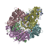 7srvSC S: Starting model for refinement C: citing same article ( |
|---|---|
| Similar structure data |
-Links
 PDBj
PDBj


- Assembly
| Deposited unit | 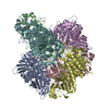
| ||||||||||||||||||||||||
|---|---|---|---|---|---|---|---|---|---|---|---|---|---|---|---|---|---|---|---|---|---|---|---|---|---|
| 1 |
| ||||||||||||||||||||||||
| Unit cell |
| ||||||||||||||||||||||||
| Noncrystallographic symmetry (NCS) | NCS oper:
|
-Components
-Protein , 1 types, 6 molecules ABCDEF
| #1: Protein | Mass: 58577.777 Da / Num. of mol.: 6 Source method: isolated from a genetically manipulated source Source: (gene. exp.) Production host:  |
|---|
-Non-polymers , 5 types, 301 molecules 


| #2: Chemical | ChemComp-CO3 /  Mass: 60.009 Da / Num. of mol.: 6 / Source method: obtained synthetically / Formula: CO3 Mass: 60.009 Da / Num. of mol.: 6 / Source method: obtained synthetically / Formula: CO3#3: Chemical | ChemComp-ZN /  Mass: 65.409 Da / Num. of mol.: 12 / Source method: obtained synthetically / Formula: Zn / Feature type: SUBJECT OF INVESTIGATION Mass: 65.409 Da / Num. of mol.: 12 / Source method: obtained synthetically / Formula: Zn / Feature type: SUBJECT OF INVESTIGATION#4: Chemical |  Mass: 118.174 Da / Num. of mol.: 2 / Source method: obtained synthetically / Formula: C6H14O2 / Comment: precipitant*YM Mass: 118.174 Da / Num. of mol.: 2 / Source method: obtained synthetically / Formula: C6H14O2 / Comment: precipitant*YM#5: Chemical |  Mass: 96.063 Da / Num. of mol.: 2 / Source method: obtained synthetically / Formula: SO4 Mass: 96.063 Da / Num. of mol.: 2 / Source method: obtained synthetically / Formula: SO4#6: Water | ChemComp-HOH / | Mass: 18.015 Da / Num. of mol.: 279 / Source method: isolated from a natural source / Formula: H2O |
|---|
-Details
| Has ligand of interest | Y |
|---|---|
| Has protein modification | N |
-Experimental details
-Experiment
| Experiment | Method: X-RAY DIFFRACTION / Number of used crystals: 1 |
|---|
- Sample preparation
| Crystal | Density Matthews: 2.49 Å3/Da / Density % sol: 50.69 % |
|---|---|
| Crystal grow | Temperature: 293 K / Method: vapor diffusion, hanging drop / Details: 50 mM HEPES pH 8.0, 150 mM NaCl |
-Data collection
| Diffraction | Mean temperature: 100 K / Serial crystal experiment: N |
|---|---|
| Diffraction source | Source: SYNCHROTRON / Site: Australian Synchrotron / Beamline: MX2 / Wavelength: 0.9537 Å |
| Detector | Type: DECTRIS EIGER X 16M / Detector: PIXEL / Date: Jul 22, 2020 |
| Radiation | Protocol: SINGLE WAVELENGTH / Monochromatic (M) / Laue (L): M / Scattering type: x-ray |
| Radiation wavelength | Wavelength: 0.9537 Å / Relative weight: 1 |
| Reflection | Resolution: 2.3→48.26 Å / Num. obs: 144874 / % possible obs: 95.49 % / Redundancy: 9.7 % / Biso Wilson estimate: 44.05 Å2 / CC1/2: 0.998 / Rmerge(I) obs: 0.1411 / Net I/σ(I): 11.81 |
| Reflection shell | Resolution: 2.3→2.385 Å / Rmerge(I) obs: 1.56 / Mean I/σ(I) obs: 1.26 / Num. unique obs: 14859 / CC1/2: 0.743 |
- Processing
| Software |
| |||||||||||||||||||||||||||||||||||||||||||||||||||||||||||||||||||||||||||||||||||||||||||||||||||||||||
|---|---|---|---|---|---|---|---|---|---|---|---|---|---|---|---|---|---|---|---|---|---|---|---|---|---|---|---|---|---|---|---|---|---|---|---|---|---|---|---|---|---|---|---|---|---|---|---|---|---|---|---|---|---|---|---|---|---|---|---|---|---|---|---|---|---|---|---|---|---|---|---|---|---|---|---|---|---|---|---|---|---|---|---|---|---|---|---|---|---|---|---|---|---|---|---|---|---|---|---|---|---|---|---|---|---|---|
| Refinement | Method to determine structure: MOLECULAR REPLACEMENT Starting model: 7SRV Resolution: 2.3→47.24 Å / SU ML: 0.3574 / Cross valid method: FREE R-VALUE / σ(F): 1.34 / Phase error: 36.6706 Stereochemistry target values: GeoStd + Monomer Library + CDL v1.2
| |||||||||||||||||||||||||||||||||||||||||||||||||||||||||||||||||||||||||||||||||||||||||||||||||||||||||
| Solvent computation | Shrinkage radii: 0.9 Å / VDW probe radii: 1.11 Å / Solvent model: FLAT BULK SOLVENT MODEL | |||||||||||||||||||||||||||||||||||||||||||||||||||||||||||||||||||||||||||||||||||||||||||||||||||||||||
| Displacement parameters | Biso mean: 57.06 Å2 | |||||||||||||||||||||||||||||||||||||||||||||||||||||||||||||||||||||||||||||||||||||||||||||||||||||||||
| Refinement step | Cycle: LAST / Resolution: 2.3→47.24 Å
| |||||||||||||||||||||||||||||||||||||||||||||||||||||||||||||||||||||||||||||||||||||||||||||||||||||||||
| Refine LS restraints |
| |||||||||||||||||||||||||||||||||||||||||||||||||||||||||||||||||||||||||||||||||||||||||||||||||||||||||
| LS refinement shell |
|
