 Movie
Movie Controller


+ Open data

- Basic information
| Entry | Database: PDB / ID: 7t2s | ||||||
|---|---|---|---|---|---|---|---|
| Title | Structure of E. coli upec-117 Cap18 3'-5' exonuclease | ||||||
 Components Components | 3'-5' exonuclease | ||||||
 Keywords Keywords | DNA BINDING PROTEIN / CBASS | ||||||
| Function / homology |  Function and homology information Function and homology informationDNA replication proofreading / 3'-5' exonuclease activity / nucleic acid binding / cytosol Similarity search - Function | ||||||
| Biological species |  | ||||||
| Method |  X-RAY DIFFRACTION / SYNCHROTRON / SAD / Resolution: 1.82 Å X-RAY DIFFRACTION / SYNCHROTRON / SAD / Resolution: 1.82 Å | ||||||
 Authors Authors | Liang, Q. / Richey, S.T. / Ye, Q. / Lau, R.K. / Corbett, K.D. | ||||||
| Funding support |  United States, 1items United States, 1items
| ||||||
 Citation Citation | Journal: Protein Sci. / Year: 2022 Title: Structure and activity of a bacterial defense-associated 3'-5' exonuclease. Authors: Liang, Q. / Richey, S.T. / Ur, S.N. / Ye, Q. / Lau, R.K. / Corbett, K.D. | ||||||
| History |
|
- Structure visualization
| Structure viewer | Molecule: MolmilJmol/JSmol |
|---|
- Downloads & links
-Download
| PDBx/mmCIF format | 7t2s.cif.gz | 138.7 KB | Display | PDBx/mmCIF format |
|---|---|---|---|---|
| PDB format | pdb7t2s.ent.gz | 92.3 KB | Display | PDB format |
| PDBx/mmJSON format | 7t2s.json.gz | Tree view | PDBx/mmJSON format | |
| Others |  Other downloads Other downloads |
-Validation report
| Arichive directory | https://data.pdbj.org/pub/pdb/validation_reports/t2/7t2sftp://data.pdbj.org/pub/pdb/validation_reports/t2/7t2s | HTTPS FTP |
|---|
-Related structure data
| Similar structure data | |
|---|---|
| Experimental dataset #1 | Data reference: 10.15785/SBGRID/862 / Data set type: diffraction image data / Details: Native data used for refinement / Metadata reference: 10.15785/SBGRID/862 |
| Experimental dataset #2 | Data reference: 10.15785/SBGRID/863 / Data set type: diffraction image data / Details: NaBr data used for phasing / Metadata reference: 10.15785/SBGRID/863 |
-Links
 PDBj
PDBj- Assembly
| Deposited unit | 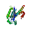
| ||||||||||||
|---|---|---|---|---|---|---|---|---|---|---|---|---|---|
| 1 | 
| ||||||||||||
| Unit cell |
| ||||||||||||
| Components on special symmetry positions |
|
-Components
| #1: Protein | Mass: 21218.156 Da / Num. of mol.: 1 Source method: isolated from a genetically manipulated source Source: (gene. exp.) |
|---|---|
| #2: Chemical | ChemComp-ACT /   Mass: 59.044 Da / Num. of mol.: 1 / Source method: obtained synthetically / Formula: C2H3O2 Mass: 59.044 Da / Num. of mol.: 1 / Source method: obtained synthetically / Formula: C2H3O2 |
| #3: Water | ChemComp-HOH /  Mass: 18.015 Da / Num. of mol.: 115 / Source method: isolated from a natural source / Formula: H2O Mass: 18.015 Da / Num. of mol.: 115 / Source method: isolated from a natural source / Formula: H2O |
| Has ligand of interest | N |
-Experimental details
-Experiment
| Experiment | Method: X-RAY DIFFRACTION / Number of used crystals: 1 |
|---|
- Sample preparation
| Crystal | Density Matthews: 3.25 Å3/Da / Density % sol: 62.1 % |
|---|---|
| Crystal grow | Temperature: 293 K / Method: vapor diffusion, hanging drop Details: 20mM Tris-HCl pH 7.5, 0.2M Li Sulfate, 0.1M Na Acetate, 0.1M HEPES pH 7.5, and 17% PEG 4000 |
-Data collection
| Diffraction | Mean temperature: 100 K / Serial crystal experiment: N | |||||||||
|---|---|---|---|---|---|---|---|---|---|---|
| Diffraction source | Source: SYNCHROTRON / Site: APS / Beamline: 24-ID-C / Wavelength: 0.9791, 0.9195 | |||||||||
| Detector | Type: DECTRIS PILATUS 6M / Detector: PIXEL / Date: Mar 28, 2019 | |||||||||
| Radiation | Protocol: SINGLE WAVELENGTH / Monochromatic (M) / Laue (L): M / Scattering type: x-ray | |||||||||
| Radiation wavelength |
| |||||||||
| Reflection | Resolution: 1.82→97.37 Å / Num. obs: 25945 / % possible obs: 99.9 % / Redundancy: 19.3 % / Biso Wilson estimate: 28.57 Å2 / CC1/2: 0.999 / Rmerge(I) obs: 0.085 / Rpim(I) all: 0.02 / Rrim(I) all: 0.088 / Net I/σ(I): 27.5 | |||||||||
| Reflection shell | Resolution: 1.82→1.86 Å / Redundancy: 18.7 % / Rmerge(I) obs: 1.039 / Mean I/σ(I) obs: 3.4 / Num. unique obs: 1491 / CC1/2: 0.868 / Rpim(I) all: 0.242 / Rrim(I) all: 1.062 / % possible all: 98.9 |
- Processing
| Software |
| ||||||||||||||||||||||||||||||||||||||||||||||||||||||||||||||||||||||
|---|---|---|---|---|---|---|---|---|---|---|---|---|---|---|---|---|---|---|---|---|---|---|---|---|---|---|---|---|---|---|---|---|---|---|---|---|---|---|---|---|---|---|---|---|---|---|---|---|---|---|---|---|---|---|---|---|---|---|---|---|---|---|---|---|---|---|---|---|---|---|---|
| Refinement | Method to determine structure: SAD / Resolution: 1.82→85.73 Å / SU ML: 0.299 / Cross valid method: FREE R-VALUE / σ(F): 0.32 / Phase error: 28.6075 Stereochemistry target values: GeoStd + Monomer Library + CDL v1.2
| ||||||||||||||||||||||||||||||||||||||||||||||||||||||||||||||||||||||
| Solvent computation | Shrinkage radii: 0.9 Å / VDW probe radii: 1.11 Å / Solvent model: FLAT BULK SOLVENT MODEL | ||||||||||||||||||||||||||||||||||||||||||||||||||||||||||||||||||||||
| Displacement parameters | Biso mean: 43.67 Å2 | ||||||||||||||||||||||||||||||||||||||||||||||||||||||||||||||||||||||
| Refinement step | Cycle: LAST / Resolution: 1.82→85.73 Å
| ||||||||||||||||||||||||||||||||||||||||||||||||||||||||||||||||||||||
| Refine LS restraints |
| ||||||||||||||||||||||||||||||||||||||||||||||||||||||||||||||||||||||
| LS refinement shell |
| ||||||||||||||||||||||||||||||||||||||||||||||||||||||||||||||||||||||
| Refinement TLS params. | Method: refined / Origin x: 25.8980420785 Å / Origin y: 44.7304485157 Å / Origin z: 50.9489171232 Å
| ||||||||||||||||||||||||||||||||||||||||||||||||||||||||||||||||||||||
| Refinement TLS group | Selection details: chain A |
