| 登録情報 | データベース: PDB / ID: 7t2q
|
|---|
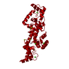
| タイトル | PEGylated Calmodulin-1 (K148U) |
|---|
 要素 要素 | Calmodulin-1 |
|---|
 キーワード キーワード | METAL BINDING PROTEIN / Calmodulin-1 |
|---|
| 機能・相同性 |  機能・相同性情報 機能・相同性情報
CaM pathway / Cam-PDE 1 activation / Sodium/Calcium exchangers / Calmodulin induced events / Reduction of cytosolic Ca++ levels / Activation of Ca-permeable Kainate Receptor / CREB1 phosphorylation through the activation of CaMKII/CaMKK/CaMKIV cascasde / Loss of phosphorylation of MECP2 at T308 / CREB1 phosphorylation through the activation of Adenylate Cyclase / CaMK IV-mediated phosphorylation of CREB ...CaM pathway / Cam-PDE 1 activation / Sodium/Calcium exchangers / Calmodulin induced events / Reduction of cytosolic Ca++ levels / Activation of Ca-permeable Kainate Receptor / CREB1 phosphorylation through the activation of CaMKII/CaMKK/CaMKIV cascasde / Loss of phosphorylation of MECP2 at T308 / CREB1 phosphorylation through the activation of Adenylate Cyclase / CaMK IV-mediated phosphorylation of CREB / PKA activation / negative regulation of high voltage-gated calcium channel activity / Glycogen breakdown (glycogenolysis) / CLEC7A (Dectin-1) induces NFAT activation / Activation of RAC1 downstream of NMDARs / negative regulation of ryanodine-sensitive calcium-release channel activity / organelle localization by membrane tethering / mitochondrion-endoplasmic reticulum membrane tethering / autophagosome membrane docking / negative regulation of calcium ion export across plasma membrane / regulation of cardiac muscle cell action potential / presynaptic endocytosis / Synthesis of IP3 and IP4 in the cytosol / regulation of cell communication by electrical coupling involved in cardiac conduction / Phase 0 - rapid depolarisation / Negative regulation of NMDA receptor-mediated neuronal transmission / calcineurin-mediated signaling / Unblocking of NMDA receptors, glutamate binding and activation / RHO GTPases activate PAKs / Ion transport by P-type ATPases / Uptake and function of anthrax toxins / regulation of ryanodine-sensitive calcium-release channel activity / Long-term potentiation / protein phosphatase activator activity / Calcineurin activates NFAT / Regulation of MECP2 expression and activity / DARPP-32 events / catalytic complex / Smooth Muscle Contraction / detection of calcium ion / regulation of cardiac muscle contraction / RHO GTPases activate IQGAPs / regulation of cardiac muscle contraction by regulation of the release of sequestered calcium ion / cellular response to interferon-beta / Protein methylation / calcium channel inhibitor activity / Activation of AMPK downstream of NMDARs / presynaptic cytosol / Ion homeostasis / regulation of release of sequestered calcium ion into cytosol by sarcoplasmic reticulum / eNOS activation / titin binding / Tetrahydrobiopterin (BH4) synthesis, recycling, salvage and regulation / sperm midpiece / regulation of calcium-mediated signaling / voltage-gated potassium channel complex / calcium channel complex / FCERI mediated Ca+2 mobilization / substantia nigra development / Ras activation upon Ca2+ influx through NMDA receptor / FCGR3A-mediated IL10 synthesis / regulation of heart rate / Antigen activates B Cell Receptor (BCR) leading to generation of second messengers / calyx of Held / adenylate cyclase activator activity / sarcomere / VEGFR2 mediated cell proliferation / protein serine/threonine kinase activator activity / regulation of cytokinesis / VEGFR2 mediated vascular permeability / spindle microtubule / calcium channel regulator activity / Translocation of SLC2A4 (GLUT4) to the plasma membrane / positive regulation of receptor signaling pathway via JAK-STAT / RAF activation / Transcriptional activation of mitochondrial biogenesis / response to calcium ion / cellular response to type II interferon / Stimuli-sensing channels / G2/M transition of mitotic cell cycle / long-term synaptic potentiation / spindle pole / RAS processing / Signaling by RAF1 mutants / calcium-dependent protein binding / Signaling by moderate kinase activity BRAF mutants / Paradoxical activation of RAF signaling by kinase inactive BRAF / Signaling downstream of RAS mutants / Signaling by BRAF and RAF1 fusions / Inactivation, recovery and regulation of the phototransduction cascade / Platelet degranulation / myelin sheath / RAF/MAP kinase cascade / Ca2+ pathway / High laminar flow shear stress activates signaling by PIEZO1 and PECAM1:CDH5:KDR in endothelial cells / vesicle / transmembrane transporter binding / Extra-nuclear estrogen signaling / G protein-coupled receptor signaling pathway / calcium ion binding類似検索 - 分子機能 EF-hand / : / Recoverin; domain 1 / EF-hand domain pair / EF-hand, calcium binding motif / EF-Hand 1, calcium-binding site / EF-hand calcium-binding domain. / EF-hand calcium-binding domain profile. / EF-hand domain / EF-hand domain pair ...EF-hand / : / Recoverin; domain 1 / EF-hand domain pair / EF-hand, calcium binding motif / EF-Hand 1, calcium-binding site / EF-hand calcium-binding domain. / EF-hand calcium-binding domain profile. / EF-hand domain / EF-hand domain pair / Orthogonal Bundle / Mainly Alpha類似検索 - ドメイン・相同性 |
|---|
| 生物種 |  Homo sapiens (ヒト) Homo sapiens (ヒト) |
|---|
| 手法 |  X線回折 / シンクロトロン / 分子置換 / 解像度: 1.95 Å X線回折 / シンクロトロン / 分子置換 / 解像度: 1.95 Å |
|---|
 データ登録者 データ登録者 | Mackay, J.P. / Payne, R.J. / Patel, K. / Dowman, L.J. |
|---|
| 資金援助 | 1件 |
|---|
 引用 引用 | ジャーナル: Nat Commun / 年: 2022
タイトル: Site-selective photocatalytic functionalization of peptides and proteins at selenocysteine.
著者: Dowman, L.J. / Kulkarni, S.S. / Alegre-Requena, J.V. / Giltrap, A.M. / Norman, A.R. / Sharma, A. / Gallegos, L.C. / Mackay, A.S. / Welegedara, A.P. / Watson, E.E. / van Raad, D. / ...著者: Dowman, L.J. / Kulkarni, S.S. / Alegre-Requena, J.V. / Giltrap, A.M. / Norman, A.R. / Sharma, A. / Gallegos, L.C. / Mackay, A.S. / Welegedara, A.P. / Watson, E.E. / van Raad, D. / Niederacher, G. / Huhmann, S. / Proschogo, N. / Patel, K. / Larance, M. / Becker, C.F.W. / Mackay, J.P. / Lakhwani, G. / Huber, T. / Paton, R.S. / Payne, R.J. |
|---|
| 履歴 | | 登録 | 2021年12月6日 | 登録サイト: RCSB / 処理サイト: RCSB |
|---|
| 改定 1.0 | 2022年10月26日 | Provider: repository / タイプ: Initial release |
|---|
| 改定 1.1 | 2023年5月17日 | Group: Database references / カテゴリ: citation / citation_author
Item: _citation.country / _citation.journal_abbrev ..._citation.country / _citation.journal_abbrev / _citation.journal_id_CSD / _citation.journal_id_ISSN / _citation.journal_volume / _citation.page_first / _citation.page_last / _citation.pdbx_database_id_DOI / _citation.pdbx_database_id_PubMed / _citation.title / _citation.year |
|---|
| 改定 1.2 | 2023年10月25日 | Group: Data collection / Refinement description
カテゴリ: chem_comp_atom / chem_comp_bond / pdbx_initial_refinement_model |
|---|
|
|---|
 ムービー
ムービー コントローラー


 データを開く
 基本情報
基本情報 構造の表示
構造の表示 ダウンロードとリンク
 その他のダウンロード
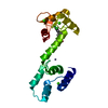
 PDBj
PDBj


 集合体
集合体


 分子量: 40.078 Da / 分子数: 4 / 由来タイプ: 合成 / 式: Ca
 分子量: 24.305 Da / 分子数: 2 / 由来タイプ: 合成 / 式: Mg
 分子量: 65.409 Da / 分子数: 1 / 由来タイプ: 天然 / 式: Zn
 分子量: 18.015 Da / 分子数: 96 / 由来タイプ: 天然 / 式: H2O
 試料調製
試料調製 / ビームライン: MX2 / 波長: 0.953736 Å
 解析
解析