| 登録情報 | データベース: PDB / ID: 7t1k
|
|---|
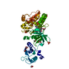
| タイトル | Crystal structure of a superbinder Fes SH2 domain (sFes1) in complex with a high affinity phosphopeptide |
|---|
 要素 要素 | - Synthetic phosphotyrosine-containing Ezrin-derived peptide
- Tyrosine-protein kinase Fes/Fps
|
|---|
 キーワード キーワード | SIGNALING PROTEIN / Src Homology 2 (SH2) / superbinder / engineered / phage display |
|---|
| 機能・相同性 |  機能・相同性情報 機能・相同性情報
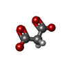
terminal web assembly / intestinal D-glucose absorption / protein localization to cell cortex / regulation of microvillus length / establishment or maintenance of apical/basal cell polarity / regulation of organelle assembly / postsynaptic actin cytoskeleton organization / positive regulation of early endosome to late endosome transport / microvillus assembly / positive regulation of myeloid cell differentiation ...terminal web assembly / intestinal D-glucose absorption / protein localization to cell cortex / regulation of microvillus length / establishment or maintenance of apical/basal cell polarity / regulation of organelle assembly / postsynaptic actin cytoskeleton organization / positive regulation of early endosome to late endosome transport / microvillus assembly / positive regulation of myeloid cell differentiation / membrane to membrane docking / Netrin-1 signaling / establishment of centrosome localization / uropod / regulation of mast cell degranulation / astral microtubule organization / negative regulation of p38MAPK cascade / positive regulation of protein localization to early endosome / protein kinase A signaling / regulation of vesicle-mediated transport / SEMA3A-Plexin repulsion signaling by inhibiting Integrin adhesion / cortical microtubule organization / cellular response to vitamin D / regulation of cell motility / filopodium assembly / CRMPs in Sema3A signaling / microtubule bundle formation / protein-containing complex localization / positive regulation of monocyte differentiation / positive regulation of multicellular organism growth / sphingosine-1-phosphate receptor signaling pathway / S100 protein binding / establishment of endothelial barrier / centrosome cycle / Sensory processing of sound by outer hair cells of the cochlea / Sensory processing of sound by inner hair cells of the cochlea / immunoglobulin receptor binding / negative regulation of interleukin-2 production / leukocyte cell-cell adhesion / negative regulation of T cell receptor signaling pathway / protein kinase A binding / microvillus membrane / cortical cytoskeleton / protein kinase A regulatory subunit binding / protein kinase A catalytic subunit binding / microvillus / Recycling pathway of L1 / brush border / regulation of cell differentiation / myoblast proliferation / actin filament bundle assembly / plasma membrane raft / immunological synapse / Sema3A PAK dependent Axon repulsion / cardiac muscle cell proliferation / regulation of cell adhesion / cell adhesion molecule binding / ruffle / positive regulation of microtubule polymerization / phosphatidylinositol binding / peptidyl-tyrosine phosphorylation / cellular response to cAMP / cell periphery / protein localization to plasma membrane / adherens junction / actin filament / cell projection / positive regulation of protein localization to plasma membrane / filopodium / non-membrane spanning protein tyrosine kinase activity / non-specific protein-tyrosine kinase / Signaling by SCF-KIT / positive regulation of neuron projection development / receptor internalization / negative regulation of ERK1 and ERK2 cascade / ruffle membrane / cytoplasmic side of plasma membrane / fibrillar center / chemotaxis / apical part of cell / positive regulation of protein catabolic process / disordered domain specific binding / actin filament binding / actin cytoskeleton / regulation of cell shape / regulation of cell population proliferation / actin binding / microtubule cytoskeleton / protein autophosphorylation / ATPase binding / actin cytoskeleton organization / cytoplasmic vesicle / protein tyrosine kinase activity / vesicle / basolateral plasma membrane / microtubule binding / endosome / cell adhesion / ciliary basal body / apical plasma membrane類似検索 - 分子機能 Tyrosine-protein kinase, Fes/Fps type / Fes/Fps/Fer, SH2 domain / Moesin tail domain superfamily / Ezrin/radixin/moesin / Ezrin/radixin/moesin, C-terminal / ERM family, FERM domain C-lobe / Ezrin/radixin/moesin, alpha-helical domain / Ezrin/radixin/moesin family C terminal / Ezrin/radixin/moesin, alpha-helical domain / Fes/CIP4, and EFC/F-BAR homology domain ...Tyrosine-protein kinase, Fes/Fps type / Fes/Fps/Fer, SH2 domain / Moesin tail domain superfamily / Ezrin/radixin/moesin / Ezrin/radixin/moesin, C-terminal / ERM family, FERM domain C-lobe / Ezrin/radixin/moesin, alpha-helical domain / Ezrin/radixin/moesin family C terminal / Ezrin/radixin/moesin, alpha-helical domain / Fes/CIP4, and EFC/F-BAR homology domain / Fes/CIP4 homology domain / FCH domain / F-BAR domain / F-BAR domain profile. / Ezrin/radixin/moesin-like / FERM, C-terminal PH-like domain / FERM C-terminal PH-like domain / FERM C-terminal PH-like domain / FERM, N-terminal / FERM N-terminal domain / FERM domain signature 1. / FERM conserved site / AH/BAR domain superfamily / FERM domain signature 2. / FERM central domain / FERM/acyl-CoA-binding protein superfamily / FERM central domain / FERM superfamily, second domain / FERM domain / FERM domain profile. / Band 4.1 domain / Band 4.1 homologues / : / SH2 domain / Src homology 2 (SH2) domain profile. / Src homology 2 domains / SH2 domain / SH2 domain superfamily / Tyrosine-protein kinase, catalytic domain / Tyrosine kinase, catalytic domain / Tyrosine protein kinases specific active-site signature. / PH-like domain superfamily / Tyrosine-protein kinase, active site / Serine-threonine/tyrosine-protein kinase, catalytic domain / Protein tyrosine and serine/threonine kinase / Ubiquitin-like domain superfamily / Protein kinase, ATP binding site / Protein kinases ATP-binding region signature. / Protein kinase domain profile. / Protein kinase domain / Protein kinase-like domain superfamily類似検索 - ドメイン・相同性 MALONATE ION / Tyrosine-protein kinase Fes/Fps / Ezrin類似検索 - 構成要素 |
|---|
| 生物種 |  Homo sapiens (ヒト) Homo sapiens (ヒト) |
|---|
| 手法 |  X線回折 / X線回折 /  シンクロトロン / シンクロトロン /  分子置換 / 解像度: 1.25 Å 分子置換 / 解像度: 1.25 Å |
|---|
 データ登録者 データ登録者 | Martyn, G.D. / Singer, A.U. / Veggiani, G. / Kurinov, I. / Sicheri, F. / Sidhu, S.S. |
|---|
| 資金援助 |  米国, 3件 米国, 3件 | 組織 | 認可番号 | 国 |
|---|
| Canadian Institutes of Health Research (CIHR) | MOP-93684 |  米国 米国 | | Canadian Institutes of Health Research (CIHR) | FDN-143277 |  米国 米国 | | National Institutes of Health/National Institute of General Medical Sciences (NIH/NIGMS) | P30 GM124165 |  米国 米国 |
|
|---|
 引用 引用 |  ジャーナル: Acs Chem.Biol. / 年: 2022 ジャーナル: Acs Chem.Biol. / 年: 2022
タイトル: Engineered SH2 Domains for Targeted Phosphoproteomics.
著者: Martyn, G.D. / Veggiani, G. / Kusebauch, U. / Morrone, S.R. / Yates, B.P. / Singer, A.U. / Tong, J. / Manczyk, N. / Gish, G. / Sun, Z. / Kurinov, I. / Sicheri, F. / Moran, M.F. / Moritz, R.L. / Sidhu, S.S. |
|---|
| 履歴 | | 登録 | 2021年12月2日 | 登録サイト: RCSB / 処理サイト: RCSB |
|---|
| 改定 1.0 | 2022年8月24日 | Provider: repository / タイプ: Initial release |
|---|
| 改定 1.1 | 2023年10月18日 | Group: Data collection / Refinement description
カテゴリ: chem_comp_atom / chem_comp_bond / pdbx_initial_refinement_model |
|---|
| 改定 1.2 | 2023年11月15日 | Group: Data collection / カテゴリ: chem_comp_atom / chem_comp_bond / Item: _chem_comp_atom.atom_id / _chem_comp_bond.atom_id_2 |
|---|
| 改定 1.3 | 2024年11月13日 | Group: Structure summary
カテゴリ: pdbx_entry_details / pdbx_modification_feature
Item: _pdbx_entry_details.has_protein_modification |
|---|
|
|---|
 データを開く
 基本情報
基本情報 要素
要素 キーワード
キーワード 機能・相同性情報
 Homo sapiens (ヒト)
 X線回折 /
  シンクロトロン /
  分子置換 / 解像度: 1.25 Å
  データ登録者
 米国, 3件
  引用
引用 ジャーナル: Acs Chem.Biol. / 年: 2022
 構造の表示
構造の表示 Molmil
 Jmol/JSmol
 ダウンロードとリンク
 ダウンロード
 7t1k.cif.gz
 PDBx/mmCIF形式
 pdb7t1k.ent.gz
 PDB形式
PDB形式 7t1k.json.gz
 PDBx/mmJSON形式
 その他のダウンロード
 7t1k_validation.pdf.gz
 wwPDB検証レポート
 7t1k_full_validation.pdf.gz
 7t1k_validation.xml.gz
 7t1k_validation.cif.gz
 https://data.pdbj.org/pub/pdb/validation_reports/t1/7t1k
 ftp://data.pdbj.org/pub/pdb/validation_reports/t1/7t1k


 F&H 検索
 リンク
リンク 集合体
集合体
 要素
要素 Homo sapiens (ヒト) / 遺伝子: FES, FPS / プラスミド: pHH1028 / 詳細 (発現宿主): Ntermimus: His6x-TEVcleavage / 発現宿主:
 Homo sapiens (ヒト) / 参照: UniProt: P15311


 X線回折 / 使用した結晶の数: 1
  試料調製
試料調製 シンクロトロン / サイト:
  APS
APS  / ビームライン: 24-ID-E / 波長: 0.97918 Å
 解析
解析 分子置換
分子置換 ムービー
ムービー コントローラー


 PDBj
PDBj


