 Movie
Movie Controller

[English] 日本語
 Yorodumi
- PDB-7t1j: Crystal structure of RUBISCO from Rhodospirillaceae bacterium BRH_c57 -
+ Open data

- Basic information
| Entry | Database: PDB / ID: 7t1j | ||||||
|---|---|---|---|---|---|---|---|
| Title | Crystal structure of RUBISCO from Rhodospirillaceae bacterium BRH_c57 | ||||||
 Components Components | Ribulose bisphosphate carboxylase | ||||||
 Keywords Keywords | LYASE / Ribulose-1 / 5-bisphosphate carboxylase-oxygenase | ||||||
| Function / homology |  Function and homology information Function and homology informationribulose-bisphosphate carboxylase / ribulose-bisphosphate carboxylase activity / reductive pentose-phosphate cycle / monooxygenase activity / magnesium ion binding Similarity search - Function | ||||||
| Biological species |  Rhodospirillaceae bacterium BRH_c57 (bacteria) Rhodospirillaceae bacterium BRH_c57 (bacteria) | ||||||
| Method |  X-RAY DIFFRACTION / SYNCHROTRON / MOLECULAR REPLACEMENT / Resolution: 1.96 Å X-RAY DIFFRACTION / SYNCHROTRON / MOLECULAR REPLACEMENT / Resolution: 1.96 Å | ||||||
 Authors Authors | Pereira, J.H. / Liu, A.K. / Shih, P.M. / Adams, P.D. | ||||||
| Funding support |  United States, 1items United States, 1items
| ||||||
 Citation Citation | Journal: Sci Adv / Year: 2022 Title: Structural plasticity enables evolution and innovation of RuBisCO assemblies. Authors: Liu, A.K. / Pereira, J.H. / Kehl, A.J. / Rosenberg, D.J. / Orr, D.J. / Chu, S.K.S. / Banda, D.M. / Hammel, M. / Adams, P.D. / Siegel, J.B. / Shih, P.M. | ||||||
| History |
|
- Structure visualization
| Structure viewer | Molecule: MolmilJmol/JSmol |
|---|
- Downloads & links
-Download
| PDBx/mmCIF format | 7t1j.cif.gz | 3 MB | Display | PDBx/mmCIF format |
|---|---|---|---|---|
| PDB format | pdb7t1j.ent.gz | 2.5 MB | Display | PDB format |
| PDBx/mmJSON format | 7t1j.json.gz | Tree view | PDBx/mmJSON format | |
| Others |  Other downloads Other downloads |
-Validation report
| Summary document | 7t1j_validation.pdf.gz | 9.4 MB | Display | wwPDB validaton report |
|---|---|---|---|---|
| Full document | 7t1j_full_validation.pdf.gz | 9.5 MB | Display | |
| Data in XML | 7t1j_validation.xml.gz | 229.1 KB | Display | |
| Data in CIF | 7t1j_validation.cif.gz | 328.8 KB | Display | |
| Arichive directory | https://data.pdbj.org/pub/pdb/validation_reports/t1/7t1jftp://data.pdbj.org/pub/pdb/validation_reports/t1/7t1j | HTTPS FTP |
-Related structure data
| Related structure data | 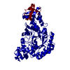 7t1cC 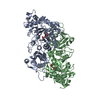 9rubS S: Starting model for refinement C: citing same article ( |
|---|---|
| Similar structure data |
-Links
 PDBj
PDBj
- Assembly
| Deposited unit | 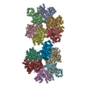
| ||||||||||||
|---|---|---|---|---|---|---|---|---|---|---|---|---|---|
| 1 | 
| ||||||||||||
| 2 | 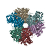
| ||||||||||||
| Unit cell |
|
-Components
| #1: Protein | Mass: 50483.938 Da / Num. of mol.: 12 Source method: isolated from a genetically manipulated source Source: (gene. exp.) Rhodospirillaceae bacterium BRH_c57 (bacteria)Gene: cbbM, VR70_18855 / Production host: References: UniProt: A0A0F2R9T6, ribulose-bisphosphate carboxylase #2: Sugar | ChemComp-CAP /   Type: saccharide / Mass: 356.115 Da / Num. of mol.: 12 / Source method: obtained synthetically / Formula: C6H14O13P2 / Feature type: SUBJECT OF INVESTIGATION Type: saccharide / Mass: 356.115 Da / Num. of mol.: 12 / Source method: obtained synthetically / Formula: C6H14O13P2 / Feature type: SUBJECT OF INVESTIGATION#3: Chemical | ChemComp-MG /   Mass: 24.305 Da / Num. of mol.: 12 / Source method: obtained synthetically / Formula: Mg / Feature type: SUBJECT OF INVESTIGATION Mass: 24.305 Da / Num. of mol.: 12 / Source method: obtained synthetically / Formula: Mg / Feature type: SUBJECT OF INVESTIGATION#4: Water | ChemComp-HOH / |  Mass: 18.015 Da / Num. of mol.: 4453 / Source method: isolated from a natural source / Formula: H2O Mass: 18.015 Da / Num. of mol.: 4453 / Source method: isolated from a natural source / Formula: H2OHas ligand of interest | Y | |
|---|
-Experimental details
-Experiment
| Experiment | Method: X-RAY DIFFRACTION / Number of used crystals: 1 |
|---|
- Sample preparation
| Crystal | Density Matthews: 2.39 Å3/Da / Density % sol: 48.58 % |
|---|---|
| Crystal grow | Temperature: 293 K / Method: vapor diffusion, sitting drop / Details: 0.2 M Magnesium Formate pH 5.9, 20 % PEG 3,350 |
-Data collection
| Diffraction | Mean temperature: 100 K / Serial crystal experiment: N |
|---|---|
| Diffraction source | Source: SYNCHROTRON / Site: NSLS / Beamline: X17B1 / Wavelength: 0.907936 Å |
| Detector | Type: DECTRIS EIGER X 16M / Detector: PIXEL / Date: Feb 27, 2021 |
| Radiation | Protocol: SINGLE WAVELENGTH / Monochromatic (M) / Laue (L): M / Scattering type: x-ray |
| Radiation wavelength | Wavelength: 0.907936 Å / Relative weight: 1 |
| Reflection | Resolution: 1.96→29.52 Å / Num. obs: 399609 / % possible obs: 98.2 % / Redundancy: 1.9 % / Biso Wilson estimate: 27.13 Å2 / CC1/2: 0.982 / Net I/σ(I): 4.81 |
| Reflection shell | Resolution: 1.96→2.03 Å / Num. unique obs: 35839 / CC1/2: 0.41 |
- Processing
| Software |
| |||||||||||||||||||||||||||||||||||||||||||||||||||||||||||||||||||||||||||||||||||||||||||||||||||||||||
|---|---|---|---|---|---|---|---|---|---|---|---|---|---|---|---|---|---|---|---|---|---|---|---|---|---|---|---|---|---|---|---|---|---|---|---|---|---|---|---|---|---|---|---|---|---|---|---|---|---|---|---|---|---|---|---|---|---|---|---|---|---|---|---|---|---|---|---|---|---|---|---|---|---|---|---|---|---|---|---|---|---|---|---|---|---|---|---|---|---|---|---|---|---|---|---|---|---|---|---|---|---|---|---|---|---|---|
| Refinement | Method to determine structure: MOLECULAR REPLACEMENT Starting model: 9rub Resolution: 1.96→29.52 Å / SU ML: 0.2501 / Cross valid method: FREE R-VALUE / σ(F): 1.35 / Phase error: 23.7648 Stereochemistry target values: GeoStd + Monomer Library + CDL v1.2
| |||||||||||||||||||||||||||||||||||||||||||||||||||||||||||||||||||||||||||||||||||||||||||||||||||||||||
| Solvent computation | Shrinkage radii: 0.9 Å / VDW probe radii: 1.11 Å / Solvent model: FLAT BULK SOLVENT MODEL | |||||||||||||||||||||||||||||||||||||||||||||||||||||||||||||||||||||||||||||||||||||||||||||||||||||||||
| Displacement parameters | Biso mean: 35.02 Å2 | |||||||||||||||||||||||||||||||||||||||||||||||||||||||||||||||||||||||||||||||||||||||||||||||||||||||||
| Refinement step | Cycle: LAST / Resolution: 1.96→29.52 Å
| |||||||||||||||||||||||||||||||||||||||||||||||||||||||||||||||||||||||||||||||||||||||||||||||||||||||||
| Refine LS restraints |
| |||||||||||||||||||||||||||||||||||||||||||||||||||||||||||||||||||||||||||||||||||||||||||||||||||||||||
| LS refinement shell |
|
