| 登録情報 | データベース: PDB / ID: 7t1j
|
|---|
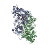
| タイトル | Crystal structure of RUBISCO from Rhodospirillaceae bacterium BRH_c57 |
|---|
 要素 要素 | Ribulose bisphosphate carboxylase |
|---|
 キーワード キーワード | LYASE / Ribulose-1 / 5-bisphosphate carboxylase-oxygenase |
|---|
| 機能・相同性 |  機能・相同性情報 機能・相同性情報
ribulose-bisphosphate carboxylase / ribulose-bisphosphate carboxylase activity / reductive pentose-phosphate cycle / monooxygenase activity / magnesium ion binding類似検索 - 分子機能 Ribulose bisphosphate carboxylase large subunit, type II / Ribulose bisphosphate carboxylase, large chain, active site / Ribulose bisphosphate carboxylase large chain active site. / Ribulose bisphosphate carboxylase, large subunit, ferrodoxin-like N-terminal / Ribulose bisphosphate carboxylase large chain, N-terminal domain / Ribulose bisphosphate carboxylase, large subunit, C-terminal / RuBisCO / Ribulose bisphosphate carboxylase, large subunit, C-terminal domain superfamily / RuBisCO large subunit, N-terminal domain superfamily / Ribulose bisphosphate carboxylase large chain, catalytic domain類似検索 - ドメイン・相同性 2-CARBOXYARABINITOL-1,5-DIPHOSPHATE / Ribulose bisphosphate carboxylase類似検索 - 構成要素 |
|---|
| 生物種 |  Rhodospirillaceae bacterium BRH_c57 (バクテリア) Rhodospirillaceae bacterium BRH_c57 (バクテリア) |
|---|
| 手法 |  X線回折 / シンクロトロン / 分子置換 / 解像度: 1.96 Å X線回折 / シンクロトロン / 分子置換 / 解像度: 1.96 Å |
|---|
 データ登録者 データ登録者 | Pereira, J.H. / Liu, A.K. / Shih, P.M. / Adams, P.D. |
|---|
| 資金援助 |  米国, 1件 米国, 1件 | 組織 | 認可番号 | 国 |
|---|
| Department of Energy (DOE, United States) | | 米国 |
|
|---|
 引用 引用 | ジャーナル: Sci Adv / 年: 2022
タイトル: Structural plasticity enables evolution and innovation of RuBisCO assemblies.
著者: Liu, A.K. / Pereira, J.H. / Kehl, A.J. / Rosenberg, D.J. / Orr, D.J. / Chu, S.K.S. / Banda, D.M. / Hammel, M. / Adams, P.D. / Siegel, J.B. / Shih, P.M. |
|---|
| 履歴 | | 登録 | 2021年12月2日 | 登録サイト: RCSB / 処理サイト: RCSB |
|---|
| 改定 1.0 | 2022年9月7日 | Provider: repository / タイプ: Initial release |
|---|
| 改定 1.1 | 2023年10月18日 | Group: Data collection / Refinement description
カテゴリ: chem_comp_atom / chem_comp_bond / pdbx_initial_refinement_model |
|---|
| 改定 1.2 | 2023年11月15日 | Group: Data collection / カテゴリ: chem_comp_atom / chem_comp_bond / Item: _chem_comp_atom.atom_id / _chem_comp_bond.atom_id_2 |
|---|
|
|---|
 ムービー
ムービー コントローラー


 データを開く
 基本情報
基本情報 構造の表示
構造の表示 ダウンロードとリンク
 その他のダウンロード
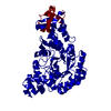
 PDBj
PDBj
 集合体
集合体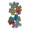

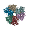

 タイプ: saccharide / 分子量: 356.115 Da / 分子数: 12 / 由来タイプ: 合成 / 式: C6H14O13P2 / タイプ: SUBJECT OF INVESTIGATION
 分子量: 24.305 Da / 分子数: 12 / 由来タイプ: 合成 / 式: Mg / タイプ: SUBJECT OF INVESTIGATION
 分子量: 18.015 Da / 分子数: 4453 / 由来タイプ: 天然 / 式: H2O
 試料調製
試料調製 解析
解析