 Movie
Movie Controller

[English] 日本語
 Yorodumi
- PDB-7t1i: Crystal structure of CAB1 Pantothenate Kinase from Saccharomyces ... -
+ Open data

- Basic information
| Entry | Database: PDB / ID: 7t1i | ||||||
|---|---|---|---|---|---|---|---|
| Title | Crystal structure of CAB1 Pantothenate Kinase from Saccharomyces cerevisiae in complex with compound YU385597 | ||||||
 Components Components | Pantothenate kinase CAB1 | ||||||
 Keywords Keywords | TRANSFERASE/TRANSFERASE INHIBITOR / kinase / TRANSFERASE / TRANSFERASE-TRANSFERASE INHIBITOR complex | ||||||
| Function / homology |  Function and homology information Function and homology informationVitamin B5 (pantothenate) metabolism / ergosterol metabolic process / pantothenate kinase / pantothenate kinase activity / coenzyme A biosynthetic process / ATP binding / nucleus / cytoplasm / cytosol Similarity search - Function | ||||||
| Biological species |  | ||||||
| Method |  X-RAY DIFFRACTION / SYNCHROTRON / MOLECULAR REPLACEMENT / Resolution: 2.4 Å X-RAY DIFFRACTION / SYNCHROTRON / MOLECULAR REPLACEMENT / Resolution: 2.4 Å | ||||||
 Authors Authors | Gihaz, S. / Ben Mamoun, C. | ||||||
| Funding support |  United States, 1items United States, 1items
| ||||||
 Citation Citation | Journal: Structure / Year: 2022 Title: High-resolution crystal structure and chemical screening reveal pantothenate kinase as a new target for antifungal development. Authors: Gihaz, S. / Gareiss, P. / Choi, J.Y. / Renard, I. / Pal, A.C. / Surovsteva, Y. / Chiu, J.E. / Thekkiniath, J. / Plummer, M. / Hungerford, W. / Montgomery, M.L. / Hosford, A. / Adams, E.M. / ...Authors: Gihaz, S. / Gareiss, P. / Choi, J.Y. / Renard, I. / Pal, A.C. / Surovsteva, Y. / Chiu, J.E. / Thekkiniath, J. / Plummer, M. / Hungerford, W. / Montgomery, M.L. / Hosford, A. / Adams, E.M. / Lightfoot, J.D. / Fox 3rd, D. / Ojo, K.K. / Staker, B.L. / Fuller, K. / Ben Mamoun, C. | ||||||
| History |
|
- Structure visualization
| Structure viewer | Molecule: MolmilJmol/JSmol |
|---|
- Downloads & links
-Download
| PDBx/mmCIF format | 7t1i.cif.gz | 147.8 KB | Display | PDBx/mmCIF format |
|---|---|---|---|---|
| PDB format | pdb7t1i.ent.gz | 112.5 KB | Display | PDB format |
| PDBx/mmJSON format | 7t1i.json.gz | Tree view | PDBx/mmJSON format | |
| Others |  Other downloads Other downloads |
-Validation report
| Arichive directory | https://data.pdbj.org/pub/pdb/validation_reports/t1/7t1iftp://data.pdbj.org/pub/pdb/validation_reports/t1/7t1i | HTTPS FTP |
|---|
-Related structure data
| Related structure data | 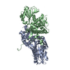 7t1gC 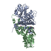 7t1hC 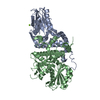 6uj5S S: Starting model for refinement C: citing same article ( |
|---|---|
| Similar structure data |
-Links
 PDBj
PDBj- Assembly
| Deposited unit | 
| ||||||||
|---|---|---|---|---|---|---|---|---|---|
| 1 |
| ||||||||
| Unit cell |
|
-Components
-Protein , 1 types, 2 molecules AB
| #1: Protein | Mass: 41974.188 Da / Num. of mol.: 2 Source method: isolated from a genetically manipulated source Source: (gene. exp.)  |
|---|
-Non-polymers , 5 types, 234 molecules 


| #2: Chemical |  Mass: 394.513 Da / Num. of mol.: 2 / Source method: obtained synthetically / Formula: C22H30N6O / Feature type: SUBJECT OF INVESTIGATION Mass: 394.513 Da / Num. of mol.: 2 / Source method: obtained synthetically / Formula: C22H30N6O / Feature type: SUBJECT OF INVESTIGATION#3: Chemical | ChemComp-SO4 /  Mass: 96.063 Da / Num. of mol.: 4 / Source method: obtained synthetically / Formula: SO4 Mass: 96.063 Da / Num. of mol.: 4 / Source method: obtained synthetically / Formula: SO4#4: Chemical |  Mass: 62.068 Da / Num. of mol.: 3 / Source method: obtained synthetically / Formula: C2H6O2 Mass: 62.068 Da / Num. of mol.: 3 / Source method: obtained synthetically / Formula: C2H6O2#5: Chemical |  Mass: 106.120 Da / Num. of mol.: 2 / Source method: obtained synthetically / Formula: C4H10O3 Mass: 106.120 Da / Num. of mol.: 2 / Source method: obtained synthetically / Formula: C4H10O3#6: Water | ChemComp-HOH / | Mass: 18.015 Da / Num. of mol.: 223 / Source method: isolated from a natural source / Formula: H2O |
|---|
-Details
| Has ligand of interest | Y |
|---|
-Experimental details
-Experiment
| Experiment | Method: X-RAY DIFFRACTION / Number of used crystals: 1 |
|---|
- Sample preparation
| Crystal | Density Matthews: 2.39 Å3/Da / Density % sol: 48.56 % |
|---|---|
| Crystal grow | Temperature: 287 K / Method: vapor diffusion, sitting drop / pH: 7 Details: 0.2 M Li Sulfate, 0.1 M Bis-Tris pH 5.5, 25% w/v PEG 3350 |
-Data collection
| Diffraction | Mean temperature: 100 K / Serial crystal experiment: N |
|---|---|
| Diffraction source | Source: SYNCHROTRON / Site: SOLEIL  / Beamline: PROXIMA 1 / Wavelength: 0.97857 Å / Beamline: PROXIMA 1 / Wavelength: 0.97857 Å |
| Detector | Type: DECTRIS EIGER X 16M / Detector: PIXEL / Date: Dec 10, 2020 |
| Radiation | Protocol: SINGLE WAVELENGTH / Monochromatic (M) / Laue (L): M / Scattering type: x-ray |
| Radiation wavelength | Wavelength: 0.97857 Å / Relative weight: 1 |
| Reflection | Resolution: 2.4→99.1 Å / Num. obs: 30602 / % possible obs: 100 % / Redundancy: 6.8 % / CC1/2: 0.994 / Rmerge(I) obs: 0.128 / Rpim(I) all: 0.05 / Net I/σ(I): 10.1 |
| Reflection shell | Resolution: 2.4→2.49 Å / Redundancy: 7 % / Rmerge(I) obs: 0.545 / Mean I/σ(I) obs: 3.7 / Num. unique obs: 2235 / CC1/2: 0.884 / Rpim(I) all: 0.216 / % possible all: 100 |
- Processing
| Software |
| |||||||||||||||||||||||||||||||||||||||||||||
|---|---|---|---|---|---|---|---|---|---|---|---|---|---|---|---|---|---|---|---|---|---|---|---|---|---|---|---|---|---|---|---|---|---|---|---|---|---|---|---|---|---|---|---|---|---|---|
| Refinement | Method to determine structure: MOLECULAR REPLACEMENT Starting model: 6UJ5 Resolution: 2.4→65.87 Å / Cor.coef. Fo:Fc: 0.948 / Cor.coef. Fo:Fc free: 0.9 / SU B: 7.045 / SU ML: 0.162 / Cross valid method: THROUGHOUT / σ(F): 0 / ESU R: 0.335 / ESU R Free: 0.237 / Stereochemistry target values: MAXIMUM LIKELIHOOD Details: HYDROGENS HAVE BEEN USED IF PRESENT IN THE INPUT U VALUES : REFINED INDIVIDUALLY
| |||||||||||||||||||||||||||||||||||||||||||||
| Solvent computation | Ion probe radii: 0.8 Å / Shrinkage radii: 0.8 Å / VDW probe radii: 1.2 Å / Solvent model: MASK | |||||||||||||||||||||||||||||||||||||||||||||
| Displacement parameters | Biso max: 136.12 Å2 / Biso mean: 35.012 Å2 / Biso min: 7.25 Å2
| |||||||||||||||||||||||||||||||||||||||||||||
| Refinement step | Cycle: final / Resolution: 2.4→65.87 Å
| |||||||||||||||||||||||||||||||||||||||||||||
| Refine LS restraints |
| |||||||||||||||||||||||||||||||||||||||||||||
| LS refinement shell | Resolution: 2.4→2.462 Å / Rfactor Rfree error: 0 / Total num. of bins used: 20
|
