 Movie
Movie Controller

[English] 日本語
 Yorodumi
- PDB-7szu: Crystal structure of Pepper RNA aptamer in complex with HBC ligan... -
+ Open data

- Basic information
| Entry | Database: PDB / ID: 7szu | ||||||
|---|---|---|---|---|---|---|---|
| Title | Crystal structure of Pepper RNA aptamer in complex with HBC ligand and Fab BL3-6 | ||||||
 Components Components |
| ||||||
 Keywords Keywords | IMMUNE SYSTEM/RNA / fluorogenic / IMMUNE SYSTEM-RNA complex | ||||||
| Function / homology | Immunoglobulins / Immunoglobulin-like / Sandwich / Mainly Beta / Chem-J8F / RNA / RNA (> 10) Function and homology information Function and homology information | ||||||
| Biological species |  synthetic construct (others) | ||||||
| Method |  X-RAY DIFFRACTION / SYNCHROTRON / MOLECULAR REPLACEMENT / Resolution: 2.24 Å X-RAY DIFFRACTION / SYNCHROTRON / MOLECULAR REPLACEMENT / Resolution: 2.24 Å | ||||||
 Authors Authors | Rees, H.C. / Piccirilli, J.A. | ||||||
| Funding support |  United States, 1items United States, 1items
| ||||||
 Citation Citation | Journal: Acs Chem.Biol. / Year: 2022 Title: Structural Basis for Fluorescence Activation by Pepper RNA. Authors: Rees, H.C. / Gogacz, W. / Li, N.S. / Koirala, D. / Piccirilli, J.A. | ||||||
| History |
|
- Structure visualization
| Structure viewer | Molecule: MolmilJmol/JSmol |
|---|
- Downloads & links
-Download
| PDBx/mmCIF format | 7szu.cif.gz | 278.7 KB | Display | PDBx/mmCIF format |
|---|---|---|---|---|
| PDB format | pdb7szu.ent.gz | 186.5 KB | Display | PDB format |
| PDBx/mmJSON format | 7szu.json.gz | Tree view | PDBx/mmJSON format | |
| Others |  Other downloads Other downloads |
-Validation report
| Arichive directory | https://data.pdbj.org/pub/pdb/validation_reports/sz/7szuftp://data.pdbj.org/pub/pdb/validation_reports/sz/7szu | HTTPS FTP |
|---|
-Related structure data
| Related structure data |  7u0yC 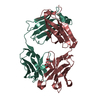 4kzdS S: Starting model for refinement C: citing same article ( |
|---|---|
| Similar structure data |
-Links
 PDBj
PDBj


- Assembly
| Deposited unit | 
| ||||||||||||
|---|---|---|---|---|---|---|---|---|---|---|---|---|---|
| 1 |
| ||||||||||||
| Unit cell |
|
-Components
-RNA chain , 1 types, 1 molecules R
| #1: RNA chain | Mass: 21734.830 Da / Num. of mol.: 1 / Source method: obtained synthetically / Source: (synth.) synthetic construct (others) |
|---|
-Antibody , 2 types, 2 molecules HL
| #2: Antibody | Mass: 23749.498 Da / Num. of mol.: 1 Source method: isolated from a genetically manipulated source Source: (gene. exp.)  |
|---|---|
| #3: Antibody | Mass: 23204.668 Da / Num. of mol.: 1 Source method: isolated from a genetically manipulated source Source: (gene. exp.) |
-Non-polymers , 4 types, 550 molecules 


| #4: Chemical |  Mass: 24.305 Da / Num. of mol.: 2 / Source method: isolated from a natural source / Formula: Mg Mass: 24.305 Da / Num. of mol.: 2 / Source method: isolated from a natural source / Formula: Mg#5: Chemical | ChemComp-J8F / |  Mass: 303.358 Da / Num. of mol.: 1 / Source method: isolated from a natural source / Formula: C19H17N3O / Feature type: SUBJECT OF INVESTIGATION Mass: 303.358 Da / Num. of mol.: 1 / Source method: isolated from a natural source / Formula: C19H17N3O / Feature type: SUBJECT OF INVESTIGATION#6: Chemical | ChemComp-NA / |  Mass: 22.990 Da / Num. of mol.: 1 / Source method: obtained synthetically / Formula: Na Mass: 22.990 Da / Num. of mol.: 1 / Source method: obtained synthetically / Formula: Na#7: Water | ChemComp-HOH / | Mass: 18.015 Da / Num. of mol.: 546 / Source method: isolated from a natural source / Formula: H2O |
|---|
-Details
| Has ligand of interest | Y |
|---|---|
| Has protein modification | Y |
-Experimental details
-Experiment
| Experiment | Method: X-RAY DIFFRACTION / Number of used crystals: 1 |
|---|
- Sample preparation
| Crystal | Density Matthews: 3.16 Å3/Da / Density % sol: 61.1 % |
|---|---|
| Crystal grow | Temperature: 298 K / Method: vapor diffusion, hanging drop / pH: 6 Details: 0.02 M Magnesium sulfate hydrate 0.002 M Cobalt(II) chloride hexahydrate 0.05 M Sodium cacodylate trihydrate pH 6.0 25% v/v (+/-)-2-Methyl-2,4-pentanediol, 0.0005 M Spermine |
-Data collection
| Diffraction | Mean temperature: 80 K / Serial crystal experiment: N |
|---|---|
| Diffraction source | Source: SYNCHROTRON / Site: APS / Beamline: 24-ID-C / Wavelength: 0.97918 Å |
| Detector | Type: DECTRIS EIGER X 16M / Detector: PIXEL / Date: Apr 10, 2021 |
| Radiation | Monochromator: M / Protocol: SINGLE WAVELENGTH / Monochromatic (M) / Laue (L): M / Scattering type: x-ray |
| Radiation wavelength | Wavelength: 0.97918 Å / Relative weight: 1 |
| Reflection | Resolution: 2.239→148.255 Å / Num. obs: 43646 / % possible obs: 99.9 % / Redundancy: 6.8 % / Biso Wilson estimate: 62.32 Å2 / CC1/2: 0.999 / Net I/σ(I): 17.9 |
| Reflection shell | Resolution: 2.24→2.31 Å / Redundancy: 6.9 % / Rmerge(I) obs: 1.449 / Mean I/σ(I) obs: 1.1 / Num. unique obs: 3937 / CC1/2: 0.605 / Rpim(I) all: 0.593 / % possible all: 99.2 |
- Processing
| Software |
| |||||||||||||||||||||||||||||||||||||||||||||||||||||||||||||||||||||||||||||||||||||||||||||||||||||||||
|---|---|---|---|---|---|---|---|---|---|---|---|---|---|---|---|---|---|---|---|---|---|---|---|---|---|---|---|---|---|---|---|---|---|---|---|---|---|---|---|---|---|---|---|---|---|---|---|---|---|---|---|---|---|---|---|---|---|---|---|---|---|---|---|---|---|---|---|---|---|---|---|---|---|---|---|---|---|---|---|---|---|---|---|---|---|---|---|---|---|---|---|---|---|---|---|---|---|---|---|---|---|---|---|---|---|---|
| Refinement | Method to determine structure: MOLECULAR REPLACEMENT Starting model: 4KZD Resolution: 2.24→58.9 Å / SU ML: 0.3874 / Cross valid method: FREE R-VALUE / σ(F): 1.34 / Phase error: 31.6424 Stereochemistry target values: GeoStd + Monomer Library + CDL v1.2
| |||||||||||||||||||||||||||||||||||||||||||||||||||||||||||||||||||||||||||||||||||||||||||||||||||||||||
| Solvent computation | Shrinkage radii: 0.9 Å / VDW probe radii: 1.11 Å / Solvent model: FLAT BULK SOLVENT MODEL | |||||||||||||||||||||||||||||||||||||||||||||||||||||||||||||||||||||||||||||||||||||||||||||||||||||||||
| Displacement parameters | Biso mean: 69.89 Å2 | |||||||||||||||||||||||||||||||||||||||||||||||||||||||||||||||||||||||||||||||||||||||||||||||||||||||||
| Refinement step | Cycle: LAST / Resolution: 2.24→58.9 Å
| |||||||||||||||||||||||||||||||||||||||||||||||||||||||||||||||||||||||||||||||||||||||||||||||||||||||||
| Refine LS restraints |
| |||||||||||||||||||||||||||||||||||||||||||||||||||||||||||||||||||||||||||||||||||||||||||||||||||||||||
| LS refinement shell |
|
