 Movie
Movie Controller


+ Open data

- Basic information
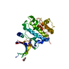
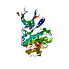
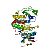
| Entry | Database: PDB / ID: 7suf | ||||||
|---|---|---|---|---|---|---|---|
| Title | Structure of CHK1 10-pt. mutant complex with LRRK2 inhibitor 06 | ||||||
 Components Components | Serine/threonine-protein kinase Chk1 | ||||||
 Keywords Keywords | TRANSFERASE/TRANSFERASE INHIBITOR / kinase / parkinson's disease / TRANSFERASE-TRANSFERASE INHIBITOR complex | ||||||
| Function / homology |  Function and homology information Function and homology informationapoptotic process involved in development / negative regulation of G0 to G1 transition / histone H3T11 kinase activity / regulation of mitotic centrosome separation / negative regulation of mitotic nuclear division / mitotic G2/M transition checkpoint / inner cell mass cell proliferation / regulation of double-strand break repair via homologous recombination / nucleus organization / negative regulation of gene expression, epigenetic ...apoptotic process involved in development / negative regulation of G0 to G1 transition / histone H3T11 kinase activity / regulation of mitotic centrosome separation / negative regulation of mitotic nuclear division / mitotic G2/M transition checkpoint / inner cell mass cell proliferation / regulation of double-strand break repair via homologous recombination / nucleus organization / negative regulation of gene expression, epigenetic / peptidyl-threonine phosphorylation / Transcriptional Regulation by E2F6 / mitotic G2 DNA damage checkpoint signaling / Presynaptic phase of homologous DNA pairing and strand exchange / replicative senescence / Activation of ATR in response to replication stress / Chk1/Chk2(Cds1) mediated inactivation of Cyclin B:Cdk1 complex / signal transduction in response to DNA damage / positive regulation of cell cycle / DNA damage checkpoint signaling / regulation of signal transduction by p53 class mediator / replication fork / condensed nuclear chromosome / TP53 Regulates Transcription of DNA Repair Genes / cellular response to mechanical stimulus / Signaling by SCF-KIT / Ubiquitin-Mediated Degradation of Phosphorylated Cdc25A / G2/M DNA damage checkpoint / G2/M transition of mitotic cell cycle / regulation of cell population proliferation / Processing of DNA double-strand break ends / Regulation of TP53 Activity through Phosphorylation / DNA replication / protein phosphorylation / protein kinase activity / non-specific serine/threonine protein kinase / chromatin remodeling / protein domain specific binding / protein serine kinase activity / DNA repair / protein serine/threonine kinase activity / apoptotic process / DNA damage response / centrosome / chromatin / protein-containing complex / extracellular space / nucleoplasm / ATP binding / nucleus / cytoplasm / cytosol Similarity search - Function | ||||||
| Biological species |  Homo sapiens (human) Homo sapiens (human) | ||||||
| Method |  X-RAY DIFFRACTION / SYNCHROTRON / MOLECULAR REPLACEMENT / Resolution: 1.48 Å X-RAY DIFFRACTION / SYNCHROTRON / MOLECULAR REPLACEMENT / Resolution: 1.48 Å | ||||||
 Authors Authors | Palte, R.L. | ||||||
| Funding support | 1items
| ||||||
 Citation Citation | Journal: J.Med.Chem. / Year: 2022 Title: Structure-Guided Discovery of Aminoquinazolines as Brain-Penetrant and Selective LRRK2 Inhibitors. Authors: Keylor, M.H. / Gulati, A. / Kattar, S.D. / Johnson, R.E. / Chau, R.W. / Margrey, K.A. / Ardolino, M.J. / Zarate, C. / Poremba, K.E. / Simov, V. / Morriello, G.J. / Acton, J.J. / Pio, B. / ...Authors: Keylor, M.H. / Gulati, A. / Kattar, S.D. / Johnson, R.E. / Chau, R.W. / Margrey, K.A. / Ardolino, M.J. / Zarate, C. / Poremba, K.E. / Simov, V. / Morriello, G.J. / Acton, J.J. / Pio, B. / Yan, X. / Palte, R.L. / McMinn, S.E. / Nogle, L. / Lesburg, C.A. / Adpressa, D. / Lin, S. / Neelamkavil, S. / Liu, P. / Su, J. / Hegde, L.G. / Woodhouse, J.D. / Faltus, R. / Xiong, T. / Ciaccio, P.J. / Piesvaux, J. / Otte, K.M. / Wood, H.B. / Kennedy, M.E. / Bennett, D.J. / DiMauro, E.F. / Fell, M.J. / Fuller, P.H. | ||||||
| History |
|
- Structure visualization
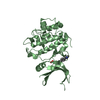
| Structure viewer | Molecule: MolmilJmol/JSmol |
|---|
- Downloads & links
-Download
| PDBx/mmCIF format | 7suf.cif.gz | 126.2 KB | Display | PDBx/mmCIF format |
|---|---|---|---|---|
| PDB format | pdb7suf.ent.gz | 95 KB | Display | PDB format |
| PDBx/mmJSON format | 7suf.json.gz | Tree view | PDBx/mmJSON format | |
| Others |  Other downloads Other downloads |
-Validation report
| Arichive directory | https://data.pdbj.org/pub/pdb/validation_reports/su/7sufftp://data.pdbj.org/pub/pdb/validation_reports/su/7suf | HTTPS FTP |
|---|
-Related structure data
| Related structure data |  7sugC  7suhC  7suiC  7sujC 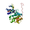 5oorS S: Starting model for refinement C: citing same article ( |
|---|---|
| Similar structure data |


-Links
 PDBj
PDBj


- Assembly
| Deposited unit | 
| ||||||||
|---|---|---|---|---|---|---|---|---|---|
| 1 |
| ||||||||
| Unit cell |
|
-Components
| #1: Protein | Mass: 34094.207 Da / Num. of mol.: 1 Mutation: N59L, V68I, L84M, Y86L, C87A, E91S, E134H, S147A, F149Y, G150S Source method: isolated from a genetically manipulated source Source: (gene. exp.) Homo sapiens (human) / Gene: CHEK1, CHK1 / Production host:   Spodoptera frugiperda (fall armyworm) Spodoptera frugiperda (fall armyworm)References: UniProt: O14757, non-specific serine/threonine protein kinase | ||||
|---|---|---|---|---|---|
| #2: Chemical | ChemComp-BVI /   Mass: 349.430 Da / Num. of mol.: 1 / Source method: obtained synthetically / Formula: C20H23N5O / Feature type: SUBJECT OF INVESTIGATION Mass: 349.430 Da / Num. of mol.: 1 / Source method: obtained synthetically / Formula: C20H23N5O / Feature type: SUBJECT OF INVESTIGATION | ||||
| #3: Chemical | ChemComp-EDO /   Mass: 62.068 Da / Num. of mol.: 4 / Source method: obtained synthetically / Formula: C2H6O2 Mass: 62.068 Da / Num. of mol.: 4 / Source method: obtained synthetically / Formula: C2H6O2#4: Water | ChemComp-HOH / |  Mass: 18.015 Da / Num. of mol.: 235 / Source method: isolated from a natural source / Formula: H2O Mass: 18.015 Da / Num. of mol.: 235 / Source method: isolated from a natural source / Formula: H2OHas ligand of interest | Y | |
-Experimental details
-Experiment
| Experiment | Method: X-RAY DIFFRACTION / Number of used crystals: 1 |
|---|
- Sample preparation
| Crystal | Density Matthews: 2.51 Å3/Da / Density % sol: 50.93 % |
|---|---|
| Crystal grow | Temperature: 293 K / Method: vapor diffusion, sitting drop / pH: 6.5 Details: 11% PEG 8000, 15-20% ethylene glycol, 0.1 M MES (pH 6.5), 5% 6-aminohexanoic acid |
-Data collection
| Diffraction | Mean temperature: 100 K / Serial crystal experiment: N |
|---|---|
| Diffraction source | Source: SYNCHROTRON / Site: ALBA  / Beamline: XALOC / Wavelength: 0.9792 Å / Beamline: XALOC / Wavelength: 0.9792 Å |
| Detector | Type: DECTRIS PILATUS 6M / Detector: PIXEL / Date: Jun 13, 2018 |
| Radiation | Protocol: SINGLE WAVELENGTH / Monochromatic (M) / Laue (L): M / Scattering type: x-ray |
| Radiation wavelength | Wavelength: 0.9792 Å / Relative weight: 1 |
| Reflection | Resolution: 1.48→57.9 Å / Num. obs: 32368 / % possible obs: 80.5 % / Redundancy: 2.9 % / CC1/2: 0.987 / Net I/σ(I): 9.4 |
| Reflection shell | Resolution: 1.483→1.61 Å / Num. unique obs: 1619 / CC1/2: 0.544 |
- Processing
| Software |
| ||||||||||||||||||||||||||||||||||||||||
|---|---|---|---|---|---|---|---|---|---|---|---|---|---|---|---|---|---|---|---|---|---|---|---|---|---|---|---|---|---|---|---|---|---|---|---|---|---|---|---|---|---|
| Refinement | Method to determine structure: MOLECULAR REPLACEMENT Starting model: 5OOR Resolution: 1.48→57.84 Å / Cor.coef. Fo:Fc: 0.938 / Cor.coef. Fo:Fc free: 0.911 / SU B: 3.927 / SU ML: 0.074 / Cross valid method: THROUGHOUT / σ(F): 0 / ESU R: 0.133 / ESU R Free: 0.129 / Stereochemistry target values: MAXIMUM LIKELIHOOD Details: HYDROGENS HAVE BEEN ADDED IN THE RIDING POSITIONS U VALUES : WITH TLS ADDED
| ||||||||||||||||||||||||||||||||||||||||
| Solvent computation | Ion probe radii: 0.8 Å / Shrinkage radii: 0.8 Å / VDW probe radii: 1.2 Å / Solvent model: MASK | ||||||||||||||||||||||||||||||||||||||||
| Displacement parameters | Biso max: 70.81 Å2 / Biso mean: 22.392 Å2 / Biso min: 8.41 Å2
| ||||||||||||||||||||||||||||||||||||||||
| Refinement step | Cycle: final / Resolution: 1.48→57.84 Å
| ||||||||||||||||||||||||||||||||||||||||
| LS refinement shell | Resolution: 1.483→1.521 Å / Rfactor Rfree error: 0 / Total num. of bins used: 20
| ||||||||||||||||||||||||||||||||||||||||
| Refinement TLS params. | Method: refined / Origin x: 5.5369 Å / Origin y: -0.1637 Å / Origin z: 6.3827 Å
|
