 Movie
Movie Controller

[English] 日本語
 Yorodumi
- PDB-7sua: Crystal Structure of the Hypothetical Protein (ACX60_00475) from ... -
+ Open data

- Basic information
| Entry | Database: PDB / ID: 7sua | ||||||
|---|---|---|---|---|---|---|---|
| Title | Crystal Structure of the Hypothetical Protein (ACX60_00475) from Acinetobacter baumannii | ||||||
 Components Components | DUF4175 domain-containing protein | ||||||
 Keywords Keywords | UNKNOWN FUNCTION / Structural Genomics / Center for Structural Genomics of Infectious Diseases / CSGID / Center for Structural Biology of Infectious Diseases / CSBID | ||||||
| Function / homology | membrane / DI(HYDROXYETHYL)ETHER / DUF4175 domain-containing protein Function and homology information Function and homology information | ||||||
| Biological species |  Acinetobacter baumannii (bacteria) Acinetobacter baumannii (bacteria) | ||||||
| Method |  X-RAY DIFFRACTION / SYNCHROTRON / SAD / Resolution: 1.65 Å X-RAY DIFFRACTION / SYNCHROTRON / SAD / Resolution: 1.65 Å | ||||||
 Authors Authors | Minasov, G. / Shuvalova, L. / Dubrovska, I. / Kiryukhina, O. / Satchell, K.J.F. / Center for Structural Biology of Infectious Diseases (CSBID) | ||||||
| Funding support |  United States, 1items United States, 1items
| ||||||
 Citation Citation | Journal: To be Published Title: Crystal Structure of the Hypothetical Protein (ACX60_00475) from Acinetobacter baumannii Authors: Minasov, G. / Shuvalova, L. / Dubrovska, I. / Kiryukhina, O. / Satchell, K.J.F. / Center for Structural Genomics of Infectious Diseases (CSGID) | ||||||
| History |
|
- Structure visualization
| Structure viewer | Molecule: MolmilJmol/JSmol |
|---|
- Downloads & links
-Download
| PDBx/mmCIF format | 7sua.cif.gz | 105.2 KB | Display | PDBx/mmCIF format |
|---|---|---|---|---|
| PDB format | pdb7sua.ent.gz | 79 KB | Display | PDB format |
| PDBx/mmJSON format | 7sua.json.gz | Tree view | PDBx/mmJSON format | |
| Others |  Other downloads Other downloads |
-Validation report
| Arichive directory | https://data.pdbj.org/pub/pdb/validation_reports/su/7suaftp://data.pdbj.org/pub/pdb/validation_reports/su/7sua | HTTPS FTP |
|---|
-Related structure data
| Similar structure data | |
|---|---|
| Other databases |
-Links
 PDBj
PDBj- Assembly
| Deposited unit | 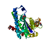
| ||||||||
|---|---|---|---|---|---|---|---|---|---|
| 1 |
| ||||||||
| Unit cell |
|
-Components
| #1: Protein | Mass: 31845.115 Da / Num. of mol.: 1 Source method: isolated from a genetically manipulated source Source: (gene. exp.) Acinetobacter baumannii (bacteria)Gene: C2U32_13765, C5H40_02315, CPI82_04650, CTZ19_00475, D8O08_001740, DLI71_12185, DLI72_04775, E1A87_06460, E2535_11650, E2539_19140, EA706_08735, EA720_011135, EA722_13985, EKS29_19110, EP550_ ...Gene: C2U32_13765, C5H40_02315, CPI82_04650, CTZ19_00475, D8O08_001740, DLI71_12185, DLI72_04775, E1A87_06460, E2535_11650, E2539_19140, EA706_08735, EA720_011135, EA722_13985, EKS29_19110, EP550_00510, EP560_17500, F2P40_11735, F4T85_08810, F4T91_09080, FE003_00465, FJU36_16290, FJU42_18320, FR761_00435, GSE42_19580, H0529_16845, HBK86_17525, IMO23_17555 Plasmid: pMCSG53 / Production host: |
|---|---|
| #2: Chemical | ChemComp-PEG /   Mass: 106.120 Da / Num. of mol.: 1 / Source method: obtained synthetically / Formula: C4H10O3 Mass: 106.120 Da / Num. of mol.: 1 / Source method: obtained synthetically / Formula: C4H10O3 |
| #3: Chemical | ChemComp-EDO /   Mass: 62.068 Da / Num. of mol.: 1 / Source method: obtained synthetically / Formula: C2H6O2 Mass: 62.068 Da / Num. of mol.: 1 / Source method: obtained synthetically / Formula: C2H6O2 |
| #4: Water | ChemComp-HOH /  Mass: 18.015 Da / Num. of mol.: 232 / Source method: isolated from a natural source / Formula: H2O Mass: 18.015 Da / Num. of mol.: 232 / Source method: isolated from a natural source / Formula: H2O |
| Has ligand of interest | N |
| Has protein modification | Y |
-Experimental details
-Experiment
| Experiment | Method: X-RAY DIFFRACTION / Number of used crystals: 1 |
|---|
- Sample preparation
| Crystal | Density Matthews: 1.8 Å3/Da / Density % sol: 31.72 % |
|---|---|
| Crystal grow | Temperature: 292 K / Method: vapor diffusion, sitting drop / pH: 6.5 Details: Protein: 8.8 mg/ml, 0.01M Tris HCl (pH 8.3); Screen: Classics II (D10), 0.1M Bis-Tris (pH 6.5), 20% (w/v) PEG5000 MME |
-Data collection
| Diffraction | Mean temperature: 100 K / Serial crystal experiment: N |
|---|---|
| Diffraction source | Source: SYNCHROTRON / Site: APS / Beamline: 21-ID-E / Wavelength: 0.97872 Å |
| Detector | Type: MARMOSAIC 300 mm CCD / Detector: CCD / Date: Mar 22, 2019 / Details: Be |
| Radiation | Monochromator: C(111) / Protocol: SINGLE WAVELENGTH / Monochromatic (M) / Laue (L): M / Scattering type: x-ray |
| Radiation wavelength | Wavelength: 0.97872 Å / Relative weight: 1 |
| Reflection | Resolution: 1.65→30 Å / Num. obs: 27958 / % possible obs: 99.5 % / Observed criterion σ(I): -3 / Redundancy: 4.3 % / Biso Wilson estimate: 22.7 Å2 / Rmerge(I) obs: 0.069 / Rpim(I) all: 0.037 / Rrim(I) all: 0.079 / Rsym value: 0.069 / Χ2: 1.572 / Net I/σ(I): 24.3 |
| Reflection shell | Resolution: 1.65→1.68 Å / Redundancy: 4.1 % / Rmerge(I) obs: 0.687 / Mean I/σ(I) obs: 2.4 / Num. unique obs: 1354 / CC1/2: 0.71 / CC star: 0.911 / Rpim(I) all: 0.379 / Rrim(I) all: 0.787 / Rsym value: 0.687 / Χ2: 1 / % possible all: 99 |
- Processing
| Software |
| |||||||||||||||||||||||||||||||||||||||||||||||||||||||||||||||||||||||||||||||||||||||||||||||||||||||||||||||||||||||||||||||||||||||||||||||||||||||||||||||||||||||||||||||
|---|---|---|---|---|---|---|---|---|---|---|---|---|---|---|---|---|---|---|---|---|---|---|---|---|---|---|---|---|---|---|---|---|---|---|---|---|---|---|---|---|---|---|---|---|---|---|---|---|---|---|---|---|---|---|---|---|---|---|---|---|---|---|---|---|---|---|---|---|---|---|---|---|---|---|---|---|---|---|---|---|---|---|---|---|---|---|---|---|---|---|---|---|---|---|---|---|---|---|---|---|---|---|---|---|---|---|---|---|---|---|---|---|---|---|---|---|---|---|---|---|---|---|---|---|---|---|---|---|---|---|---|---|---|---|---|---|---|---|---|---|---|---|---|---|---|---|---|---|---|---|---|---|---|---|---|---|---|---|---|---|---|---|---|---|---|---|---|---|---|---|---|---|---|---|---|---|
| Refinement | Method to determine structure: SAD / Resolution: 1.65→27.06 Å / Cor.coef. Fo:Fc: 0.964 / Cor.coef. Fo:Fc free: 0.945 / SU B: 3.616 / SU ML: 0.061 / Cross valid method: THROUGHOUT / σ(F): 0 / ESU R: 0.095 / ESU R Free: 0.095 / Stereochemistry target values: MAXIMUM LIKELIHOOD Details: HYDROGENS HAVE BEEN ADDED IN THE RIDING POSITIONS U VALUES : WITH TLS ADDED
| |||||||||||||||||||||||||||||||||||||||||||||||||||||||||||||||||||||||||||||||||||||||||||||||||||||||||||||||||||||||||||||||||||||||||||||||||||||||||||||||||||||||||||||||
| Solvent computation | Ion probe radii: 0.8 Å / Shrinkage radii: 0.8 Å / VDW probe radii: 1.2 Å / Solvent model: MASK | |||||||||||||||||||||||||||||||||||||||||||||||||||||||||||||||||||||||||||||||||||||||||||||||||||||||||||||||||||||||||||||||||||||||||||||||||||||||||||||||||||||||||||||||
| Displacement parameters | Biso max: 98.08 Å2 / Biso mean: 25.796 Å2 / Biso min: 13.01 Å2
| |||||||||||||||||||||||||||||||||||||||||||||||||||||||||||||||||||||||||||||||||||||||||||||||||||||||||||||||||||||||||||||||||||||||||||||||||||||||||||||||||||||||||||||||
| Refinement step | Cycle: final / Resolution: 1.65→27.06 Å
| |||||||||||||||||||||||||||||||||||||||||||||||||||||||||||||||||||||||||||||||||||||||||||||||||||||||||||||||||||||||||||||||||||||||||||||||||||||||||||||||||||||||||||||||
| Refine LS restraints |
| |||||||||||||||||||||||||||||||||||||||||||||||||||||||||||||||||||||||||||||||||||||||||||||||||||||||||||||||||||||||||||||||||||||||||||||||||||||||||||||||||||||||||||||||
| LS refinement shell | Resolution: 1.655→1.697 Å / Rfactor Rfree error: 0 / Total num. of bins used: 20
| |||||||||||||||||||||||||||||||||||||||||||||||||||||||||||||||||||||||||||||||||||||||||||||||||||||||||||||||||||||||||||||||||||||||||||||||||||||||||||||||||||||||||||||||
| Refinement TLS params. | Method: refined / Refine-ID: X-RAY DIFFRACTION
| |||||||||||||||||||||||||||||||||||||||||||||||||||||||||||||||||||||||||||||||||||||||||||||||||||||||||||||||||||||||||||||||||||||||||||||||||||||||||||||||||||||||||||||||
| Refinement TLS group |
|
