 Movie
Movie Controller

[English] 日本語
 Yorodumi
- PDB-7su8: DIHYDRONEOPTERIN ALDOLASE (DHNA) FROM YERSINIA PESTIS with alkyla... -
+ Open data

- Basic information
| Entry | Database: PDB / ID: 7su8 | ||||||
|---|---|---|---|---|---|---|---|
| Title | DIHYDRONEOPTERIN ALDOLASE (DHNA) FROM YERSINIA PESTIS with alkylated Cys50 CO-CRYSTALLIZED with 7,8-dihydroneopterin | ||||||
 Components Components | 7,8-dihydroneopterin aldolase | ||||||
 Keywords Keywords | BIOSYNTHETIC PROTEIN / FOLB FOLATE PATHWAY | ||||||
| Function / homology |  Function and homology information Function and homology informationdihydroneopterin aldolase / dihydroneopterin aldolase activity / folic acid biosynthetic process / isomerase activity / tetrahydrofolate biosynthetic process / kinase activity Similarity search - Function | ||||||
| Biological species |   Yersinia pestis (bacteria) Yersinia pestis (bacteria) | ||||||
| Method |  X-RAY DIFFRACTION / MOLECULAR REPLACEMENT / Resolution: 2.65 Å X-RAY DIFFRACTION / MOLECULAR REPLACEMENT / Resolution: 2.65 Å | ||||||
 Authors Authors | Bourne, C.R. | ||||||
| Funding support | 1items
| ||||||
 Citation Citation | Journal: To Be Published Title: DIHYDRONEOPTERIN ALDOLASE (DHNA) FROM YERSINIA PESTIS with alkylated Cys50 CO-CRYSTALLIZED with 7,8-dihydroneopterin Authors: Bourne, C.R. | ||||||
| History |
|
- Structure visualization
| Structure viewer | Molecule: MolmilJmol/JSmol |
|---|
- Downloads & links
-Download
| PDBx/mmCIF format | 7su8.cif.gz | 66.1 KB | Display | PDBx/mmCIF format |
|---|---|---|---|---|
| PDB format | pdb7su8.ent.gz | 46.1 KB | Display | PDB format |
| PDBx/mmJSON format | 7su8.json.gz | Tree view | PDBx/mmJSON format | |
| Others |  Other downloads Other downloads |
-Validation report
| Arichive directory | https://data.pdbj.org/pub/pdb/validation_reports/su/7su8ftp://data.pdbj.org/pub/pdb/validation_reports/su/7su8 | HTTPS FTP |
|---|
-Related structure data
| Related structure data |  6ojoS S: Starting model for refinement |
|---|---|
| Similar structure data |
-Links
 PDBj
PDBj
- Assembly
| Deposited unit | 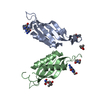
| ||||||||
|---|---|---|---|---|---|---|---|---|---|
| 1 | 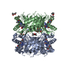
| ||||||||
| Unit cell |
|
-Components
-Protein , 1 types, 2 molecules AB
| #1: Protein | Mass: 16149.166 Da / Num. of mol.: 2 Source method: isolated from a genetically manipulated source Source: (gene. exp.) Yersinia pestis (bacteria) / Gene: y3531 / Plasmid: pMCSG7 / Production host: |
|---|
-Non-polymers , 5 types, 67 molecules 


| #2: Chemical |  Mass: 127.141 Da / Num. of mol.: 2 / Source method: obtained synthetically / Formula: C6H9NO2 / Feature type: SUBJECT OF INVESTIGATION Mass: 127.141 Da / Num. of mol.: 2 / Source method: obtained synthetically / Formula: C6H9NO2 / Feature type: SUBJECT OF INVESTIGATION#3: Chemical |  Mass: 195.179 Da / Num. of mol.: 2 / Source method: obtained synthetically / Formula: C7H9N5O2 / Feature type: SUBJECT OF INVESTIGATION Mass: 195.179 Da / Num. of mol.: 2 / Source method: obtained synthetically / Formula: C7H9N5O2 / Feature type: SUBJECT OF INVESTIGATION#4: Chemical |  Mass: 118.174 Da / Num. of mol.: 2 / Source method: obtained synthetically / Formula: C6H14O2 / Comment: precipitant*YM Mass: 118.174 Da / Num. of mol.: 2 / Source method: obtained synthetically / Formula: C6H14O2 / Comment: precipitant*YM#5: Chemical | ChemComp-TRS / |  Mass: 122.143 Da / Num. of mol.: 1 / Source method: obtained synthetically / Formula: C4H12NO3 / Comment: pH buffer*YM Mass: 122.143 Da / Num. of mol.: 1 / Source method: obtained synthetically / Formula: C4H12NO3 / Comment: pH buffer*YM#6: Water | ChemComp-HOH / | Mass: 18.015 Da / Num. of mol.: 60 / Source method: isolated from a natural source / Formula: H2O |
|---|
-Details
| Has ligand of interest | Y |
|---|---|
| Has protein modification | Y |
-Experimental details
-Experiment
| Experiment | Method: X-RAY DIFFRACTION / Number of used crystals: 1 |
|---|
- Sample preparation
| Crystal | Density Matthews: 2.16 Å3/Da / Density % sol: 43.1 % |
|---|---|
| Crystal grow | Temperature: 293 K / Method: vapor diffusion, sitting drop Details: 0.05 - 0.1M BisTris, 0.1-0.2 M CaCl2, 0 - 0.1M NaCl, 43 - 49% MPD PH range: 6.2 - 6.7 |
-Data collection
| Diffraction | Mean temperature: 100 K / Serial crystal experiment: N | |||||||||||||||||||||||||||||||||||||||||||||||||||||||||||||||||||||||||||||||||||||||||||||||||||||||||||||||||||||||||||||||||||||||||||||||||||||||||||||||||||||||||||||||||||||||||||||
|---|---|---|---|---|---|---|---|---|---|---|---|---|---|---|---|---|---|---|---|---|---|---|---|---|---|---|---|---|---|---|---|---|---|---|---|---|---|---|---|---|---|---|---|---|---|---|---|---|---|---|---|---|---|---|---|---|---|---|---|---|---|---|---|---|---|---|---|---|---|---|---|---|---|---|---|---|---|---|---|---|---|---|---|---|---|---|---|---|---|---|---|---|---|---|---|---|---|---|---|---|---|---|---|---|---|---|---|---|---|---|---|---|---|---|---|---|---|---|---|---|---|---|---|---|---|---|---|---|---|---|---|---|---|---|---|---|---|---|---|---|---|---|---|---|---|---|---|---|---|---|---|---|---|---|---|---|---|---|---|---|---|---|---|---|---|---|---|---|---|---|---|---|---|---|---|---|---|---|---|---|---|---|---|---|---|---|---|---|---|---|
| Diffraction source | Source: ROTATING ANODE / Type: RIGAKU MICROMAX-007 HF / Wavelength: 1.54 Å | |||||||||||||||||||||||||||||||||||||||||||||||||||||||||||||||||||||||||||||||||||||||||||||||||||||||||||||||||||||||||||||||||||||||||||||||||||||||||||||||||||||||||||||||||||||||||||||
| Detector | Type: DECTRIS PILATUS 200K / Detector: PIXEL / Date: May 31, 2021 | |||||||||||||||||||||||||||||||||||||||||||||||||||||||||||||||||||||||||||||||||||||||||||||||||||||||||||||||||||||||||||||||||||||||||||||||||||||||||||||||||||||||||||||||||||||||||||||
| Radiation | Protocol: SINGLE WAVELENGTH / Monochromatic (M) / Laue (L): M / Scattering type: x-ray | |||||||||||||||||||||||||||||||||||||||||||||||||||||||||||||||||||||||||||||||||||||||||||||||||||||||||||||||||||||||||||||||||||||||||||||||||||||||||||||||||||||||||||||||||||||||||||||
| Radiation wavelength | Wavelength: 1.54 Å / Relative weight: 1 | |||||||||||||||||||||||||||||||||||||||||||||||||||||||||||||||||||||||||||||||||||||||||||||||||||||||||||||||||||||||||||||||||||||||||||||||||||||||||||||||||||||||||||||||||||||||||||||
| Reflection | Resolution: 2.65→50 Å / Num. obs: 7332 / % possible obs: 91.8 % / Redundancy: 3.5 % / Rmerge(I) obs: 0.043 / Rpim(I) all: 0.025 / Rrim(I) all: 0.05 / Χ2: 0.55 / Net I/σ(I): 9.7 / Num. measured all: 25781 | |||||||||||||||||||||||||||||||||||||||||||||||||||||||||||||||||||||||||||||||||||||||||||||||||||||||||||||||||||||||||||||||||||||||||||||||||||||||||||||||||||||||||||||||||||||||||||||
| Reflection shell | Diffraction-ID: 1
|
- Processing
| Software |
| ||||||||||||||||||||||||||||||||||||||||||
|---|---|---|---|---|---|---|---|---|---|---|---|---|---|---|---|---|---|---|---|---|---|---|---|---|---|---|---|---|---|---|---|---|---|---|---|---|---|---|---|---|---|---|---|
| Refinement | Method to determine structure: MOLECULAR REPLACEMENT Starting model: 6ojo Resolution: 2.65→36.87 Å / SU ML: 0.35 / Cross valid method: THROUGHOUT / σ(F): 1.34 / Phase error: 28.89 / Stereochemistry target values: ML
| ||||||||||||||||||||||||||||||||||||||||||
| Solvent computation | Shrinkage radii: 0.9 Å / VDW probe radii: 1.11 Å / Solvent model: FLAT BULK SOLVENT MODEL | ||||||||||||||||||||||||||||||||||||||||||
| Displacement parameters | Biso max: 117.37 Å2 / Biso mean: 53.2409 Å2 / Biso min: 19.89 Å2 | ||||||||||||||||||||||||||||||||||||||||||
| Refinement step | Cycle: final / Resolution: 2.65→36.87 Å
| ||||||||||||||||||||||||||||||||||||||||||
| Refine LS restraints |
| ||||||||||||||||||||||||||||||||||||||||||
| LS refinement shell | Refine-ID: X-RAY DIFFRACTION / Rfactor Rfree error: 0 / Total num. of bins used: 5
|
