 Movie
Movie Controller

[English] 日本語
 Yorodumi
- PDB-7sqw: Structure of the KcsA-W67F mutant with the activation gate in the... -
+ Open data

- Basic information
| Entry | Database: PDB / ID: 7sqw | |||||||||
|---|---|---|---|---|---|---|---|---|---|---|
| Title | Structure of the KcsA-W67F mutant with the activation gate in the closed conformation | |||||||||
 Components Components |
| |||||||||
 Keywords Keywords | MEMBRANE PROTEIN / channel / Fab complex / C-type inactivation | |||||||||
| Function / homology | Immunoglobulins / Immunoglobulin-like / Sandwich / Mainly Beta / Chem-1EM / NONAN-1-OL / :  Function and homology information Function and homology information | |||||||||
| Biological species |   Streptomyces (bacteria) Streptomyces (bacteria) | |||||||||
| Method |  X-RAY DIFFRACTION / SYNCHROTRON / MOLECULAR REPLACEMENT / molecular replacement / Resolution: 3.21 Å X-RAY DIFFRACTION / SYNCHROTRON / MOLECULAR REPLACEMENT / molecular replacement / Resolution: 3.21 Å | |||||||||
 Authors Authors | Cuello, L.G. / Labro, A.J. | |||||||||
| Funding support |  United States, United States,  Belgium, 2items Belgium, 2items
| |||||||||
 Citation Citation | Journal: Sci Adv / Year: 2022 Title: The nonconducting W434F mutant adopts upon membrane depolarization an inactivated-like state that differs from wild-type Shaker-IR potassium channels. Authors: Coonen, L. / Martinez-Morales, E. / Van De Sande, D.V. / Snyders, D.J. / Cortes, D.M. / Cuello, L.G. / Labro, A.J. | |||||||||
| History |
|
- Structure visualization
| Structure viewer | Molecule: MolmilJmol/JSmol |
|---|
- Downloads & links
-Download
| PDBx/mmCIF format | 7sqw.cif.gz | 217 KB | Display | PDBx/mmCIF format |
|---|---|---|---|---|
| PDB format | pdb7sqw.ent.gz | 174.5 KB | Display | PDB format |
| PDBx/mmJSON format | 7sqw.json.gz | Tree view | PDBx/mmJSON format | |
| Others |  Other downloads Other downloads |
-Validation report
| Summary document | 7sqw_validation.pdf.gz | 666.9 KB | Display | wwPDB validaton report |
|---|---|---|---|---|
| Full document | 7sqw_full_validation.pdf.gz | 676.4 KB | Display | |
| Data in XML | 7sqw_validation.xml.gz | 21.4 KB | Display | |
| Data in CIF | 7sqw_validation.cif.gz | 28.3 KB | Display | |
| Arichive directory | https://data.pdbj.org/pub/pdb/validation_reports/sq/7sqwftp://data.pdbj.org/pub/pdb/validation_reports/sq/7sqw | HTTPS FTP |
-Related structure data
| Related structure data |  1k4cS S: Starting model for refinement |
|---|---|
| Similar structure data |
-Links
 PDBj
PDBj


- Assembly
| Deposited unit | 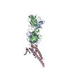
| ||||||||||||||||||
|---|---|---|---|---|---|---|---|---|---|---|---|---|---|---|---|---|---|---|---|
| 1 |
| ||||||||||||||||||
| Unit cell |
| ||||||||||||||||||
| Components on special symmetry positions |
|
-Components
-Protein , 1 types, 1 molecules C
| #3: Protein | Mass: 10939.700 Da / Num. of mol.: 1 / Mutation: W67F Source method: isolated from a genetically manipulated source Source: (gene. exp.) Streptomyces (bacteria) / Production host: Streptomyces (bacteria) |
|---|
-Antibody , 2 types, 2 molecules AB
| #1: Antibody | Mass: 23411.242 Da / Num. of mol.: 1 Source method: isolated from a genetically manipulated source Source: (gene. exp.) |
|---|---|
| #2: Antibody | Mass: 23435.738 Da / Num. of mol.: 1 Source method: isolated from a genetically manipulated source Source: (gene. exp.) |
-Non-polymers , 3 types, 7 molecules 


| #4: Chemical | ChemComp-F09 /  Mass: 144.254 Da / Num. of mol.: 1 / Source method: obtained synthetically / Formula: C9H20O Mass: 144.254 Da / Num. of mol.: 1 / Source method: obtained synthetically / Formula: C9H20O |
|---|---|
| #5: Chemical | ChemComp-1EM / ( Mass: 442.672 Da / Num. of mol.: 1 / Source method: obtained synthetically / Formula: C26H50O5 / Feature type: SUBJECT OF INVESTIGATION Mass: 442.672 Da / Num. of mol.: 1 / Source method: obtained synthetically / Formula: C26H50O5 / Feature type: SUBJECT OF INVESTIGATION |
| #6: Chemical | ChemComp-K /  Mass: 39.098 Da / Num. of mol.: 5 / Source method: obtained synthetically / Formula: K Mass: 39.098 Da / Num. of mol.: 5 / Source method: obtained synthetically / Formula: K |
-Details
| Has ligand of interest | Y |
|---|---|
| Has protein modification | Y |
-Experimental details
-Experiment
| Experiment | Method: X-RAY DIFFRACTION / Number of used crystals: 1 |
|---|
- Sample preparation
| Crystal | Density Matthews: 3.89 Å3/Da / Density % sol: 68.36 % |
|---|---|
| Crystal grow | Temperature: 292 K / Method: vapor diffusion, sitting drop Details: PEG400 , 50 mM magnesium acetate, 50 mM sodium acetate, 300 mM KCl |
-Data collection
| Diffraction | Mean temperature: 100 K / Serial crystal experiment: N |
|---|---|
| Diffraction source | Source: SYNCHROTRON / Site: SSRL / Beamline: BL14-1 / Wavelength: 1.2 Å |
| Detector | Type: DECTRIS PILATUS 6M / Detector: PIXEL / Date: Sep 19, 2014 |
| Radiation | Protocol: SINGLE WAVELENGTH / Monochromatic (M) / Laue (L): M / Scattering type: x-ray |
| Radiation wavelength | Wavelength: 1.2 Å / Relative weight: 1 |
| Reflection | Resolution: 3.21→48.69 Å / Num. obs: 14014 / % possible obs: 99.8 % / Redundancy: 3.1 % / Biso Wilson estimate: 81.33 Å2 / CC1/2: 0.75 / Net I/σ(I): 14.7 |
| Reflection shell | Resolution: 3.21→48.69 Å / Num. unique obs: 14012 / CC1/2: 0.5 / % possible all: 99.8 |
-Phasing
| Phasing | Method: molecular replacement |
|---|
- Processing
| Software |
| |||||||||||||||||||||||||||||||||||||||||||||||||||||||||||||||||||||||||||||
|---|---|---|---|---|---|---|---|---|---|---|---|---|---|---|---|---|---|---|---|---|---|---|---|---|---|---|---|---|---|---|---|---|---|---|---|---|---|---|---|---|---|---|---|---|---|---|---|---|---|---|---|---|---|---|---|---|---|---|---|---|---|---|---|---|---|---|---|---|---|---|---|---|---|---|---|---|---|---|
| Refinement | Method to determine structure: MOLECULAR REPLACEMENT Starting model: 1K4C Resolution: 3.21→48.69 Å / SU ML: 0.46 / Cross valid method: THROUGHOUT / σ(F): 1.34 / Phase error: 30.67 / Stereochemistry target values: ML
| |||||||||||||||||||||||||||||||||||||||||||||||||||||||||||||||||||||||||||||
| Solvent computation | Shrinkage radii: 0.9 Å / VDW probe radii: 1.11 Å / Solvent model: FLAT BULK SOLVENT MODEL | |||||||||||||||||||||||||||||||||||||||||||||||||||||||||||||||||||||||||||||
| Displacement parameters | Biso max: 165.2 Å2 / Biso mean: 89.551 Å2 / Biso min: 34.18 Å2 | |||||||||||||||||||||||||||||||||||||||||||||||||||||||||||||||||||||||||||||
| Refinement step | Cycle: final / Resolution: 3.21→48.69 Å
| |||||||||||||||||||||||||||||||||||||||||||||||||||||||||||||||||||||||||||||
| LS refinement shell | Refine-ID: X-RAY DIFFRACTION / Rfactor Rfree error: 0 / Total num. of bins used: 10
| |||||||||||||||||||||||||||||||||||||||||||||||||||||||||||||||||||||||||||||
| Refinement TLS params. | Method: refined / Origin x: -27.6765 Å / Origin y: -19.5068 Å / Origin z: -27.4419 Å
| |||||||||||||||||||||||||||||||||||||||||||||||||||||||||||||||||||||||||||||
| Refinement TLS group |
|
