 Movie
Movie Controller


+ Open data

- Basic information
| Entry | Database: PDB / ID: 7sqk | ||||||
|---|---|---|---|---|---|---|---|
| Title | Cryo-EM structure of the human augmin complex | ||||||
 Components Components |
| ||||||
 Keywords Keywords | CELL CYCLE / Cell Division / Mitosis / Microtubules | ||||||
| Function / homology |  Function and homology information Function and homology informationHAUS complex / regulation of microtubule nucleation / microtubule minus-end binding / nuclear microtubule / microtubule organizing center organization / mitotic spindle microtubule / microtubule nucleation / thioesterase binding / centrosome cycle / microtubule organizing center ...HAUS complex / regulation of microtubule nucleation / microtubule minus-end binding / nuclear microtubule / microtubule organizing center organization / mitotic spindle microtubule / microtubule nucleation / thioesterase binding / centrosome cycle / microtubule organizing center / intercellular bridge / spindle assembly / Loss of Nlp from mitotic centrosomes / Loss of proteins required for interphase microtubule organization from the centrosome / Recruitment of mitotic centrosome proteins and complexes / Recruitment of NuMA to mitotic centrosomes / Anchoring of the basal body to the plasma membrane / AURKA Activation by TPX2 / microtubule cytoskeleton organization / spindle pole / mitotic spindle / Regulation of PLK1 Activity at G2/M Transition / sperm principal piece / microtubule cytoskeleton / sperm midpiece / microtubule binding / cilium / ciliary basal body / cell division / centrosome / nucleolus / mitochondrion / nucleoplasm / plasma membrane / cytoplasm / cytosol Similarity search - Function | ||||||
| Biological species |  Homo sapiens (human) Homo sapiens (human) | ||||||
| Method | ELECTRON MICROSCOPY / single particle reconstruction / cryo EM / Resolution: 8 Å | ||||||
 Authors Authors | Gabel, C.A. / Chang, L. | ||||||
| Funding support | 1items
| ||||||
 Citation Citation | Journal: Nat Commun / Year: 2022 Title: Molecular architecture of the augmin complex. Authors: Clinton A Gabel / Zhuang Li / Andrew G DeMarco / Ziguo Zhang / Jing Yang / Mark C Hall / David Barford / Leifu Chang /   Abstract: Accurate segregation of chromosomes during mitosis depends on the correct assembly of the mitotic spindle, a bipolar structure composed mainly of microtubules. The augmin complex, or homologous to ...Accurate segregation of chromosomes during mitosis depends on the correct assembly of the mitotic spindle, a bipolar structure composed mainly of microtubules. The augmin complex, or homologous to augmin subunits (HAUS) complex, is an eight-subunit protein complex required for building robust mitotic spindles in metazoa. Augmin increases microtubule density within the spindle by recruiting the γ-tubulin ring complex (γ-TuRC) to pre-existing microtubules and nucleating branching microtubules. Here, we elucidate the molecular architecture of augmin by single particle cryo-electron microscopy (cryo-EM), computational methods, and crosslinking mass spectrometry (CLMS). Augmin's highly flexible structure contains a V-shaped head and a filamentous tail, with the head existing in either extended or contracted conformational states. Our work highlights how cryo-EM, complemented by computational advances and CLMS, can elucidate the structure of a challenging protein complex and provides insights into the function of augmin in mediating microtubule branching nucleation. | ||||||
| History |
|
- Structure visualization
| Structure viewer | Molecule: MolmilJmol/JSmol |
|---|
- Downloads & links
-Download
| PDBx/mmCIF format | 7sqk.cif.gz | 534.7 KB | Display | PDBx/mmCIF format |
|---|---|---|---|---|
| PDB format | pdb7sqk.ent.gz | 434 KB | Display | PDB format |
| PDBx/mmJSON format | 7sqk.json.gz | Tree view | PDBx/mmJSON format | |
| Others |  Other downloads Other downloads |
-Validation report
| Arichive directory | https://data.pdbj.org/pub/pdb/validation_reports/sq/7sqkftp://data.pdbj.org/pub/pdb/validation_reports/sq/7sqk | HTTPS FTP |
|---|
-Related structure data
| Related structure data |  25387MC M: map data used to model this data C: citing same article ( |
|---|---|
| Similar structure data |
-Links
 PDBj
PDBj
- Assembly
| Deposited unit | 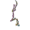
|
|---|---|
| 1 |
|
-Components
-HAUS augmin-like complex subunit ... , 7 types, 7 molecules ABCEFGH
| #1: Protein | Mass: 31910.348 Da / Num. of mol.: 1 Source method: isolated from a genetically manipulated source Source: (gene. exp.) Homo sapiens (human) / Gene: HAUS1, CCDC5, HEIC / Production host:  Trichoplusia ni (cabbage looper) / References: UniProt: Q96CS2 Trichoplusia ni (cabbage looper) / References: UniProt: Q96CS2 |
|---|---|
| #2: Protein | Mass: 32487.453 Da / Num. of mol.: 1 Source method: isolated from a genetically manipulated source Source: (gene. exp.) Homo sapiens (human) / Gene: HAUS2, C15orf25, CEP27 / Production host: Trichoplusia ni (cabbage looper) / References: UniProt: Q9NVX0 |
| #3: Protein | Mass: 69740.180 Da / Num. of mol.: 1 Source method: isolated from a genetically manipulated source Source: (gene. exp.) Homo sapiens (human) / Gene: HAUS3, C4orf15 / Production host: Trichoplusia ni (cabbage looper) / References: UniProt: Q68CZ6 |
| #5: Protein | Mass: 71783.406 Da / Num. of mol.: 1 Source method: isolated from a genetically manipulated source Source: (gene. exp.) Homo sapiens (human) / Gene: HAUS5, KIAA0841 / Production host: Trichoplusia ni (cabbage looper) / References: UniProt: O94927 |
| #6: Protein | Mass: 50250.039 Da / Num. of mol.: 1 Source method: isolated from a genetically manipulated source Source: (gene. exp.) Homo sapiens (human) / Gene: HAUS6, DGT6, FAM29A, KIAA1574 / Production host: Trichoplusia ni (cabbage looper) / References: UniProt: Q7Z4H7 |
| #7: Protein | Mass: 40819.152 Da / Num. of mol.: 1 Source method: isolated from a genetically manipulated source Source: (gene. exp.) Homo sapiens (human) / Gene: HAUS7, UCHL5IP, UIP1 / Production host: Trichoplusia ni (cabbage looper) / References: UniProt: Q99871 |
| #8: Protein | Mass: 44922.430 Da / Num. of mol.: 1 Source method: isolated from a genetically manipulated source Source: (gene. exp.) Homo sapiens (human) / Gene: HAUS8, HICE1 / Production host: Trichoplusia ni (cabbage looper) / References: UniProt: Q9BT25 |
-Protein , 1 types, 1 molecules D
| #4: Protein | Mass: 37269.805 Da / Num. of mol.: 1 Source method: isolated from a genetically manipulated source Source: (gene. exp.) Homo sapiens (human) / Gene: HAUS4, C14orf94 / Production host: Trichoplusia ni (cabbage looper) / References: UniProt: Q9H6D7 |
|---|
-Experimental details
-Experiment
| Experiment | Method: ELECTRON MICROSCOPY |
|---|---|
| EM experiment | Aggregation state: PARTICLE / 3D reconstruction method: single particle reconstruction |
- Sample preparation
| Component | Name: Holocomplex of the Homologous to Augment Subunits Complex Type: COMPLEX / Entity ID: all / Source: RECOMBINANT | |||||||||||||||||||||||||
|---|---|---|---|---|---|---|---|---|---|---|---|---|---|---|---|---|---|---|---|---|---|---|---|---|---|---|
| Molecular weight | Value: 378 kDa/nm / Experimental value: NO | |||||||||||||||||||||||||
| Source (natural) | Organism: Homo sapiens (human) | |||||||||||||||||||||||||
| Source (recombinant) | Organism: Trichoplusia ni (cabbage looper) | |||||||||||||||||||||||||
| Buffer solution | pH: 8 | |||||||||||||||||||||||||
| Buffer component |
| |||||||||||||||||||||||||
| Specimen | Conc.: 0.05 mg/ml / Embedding applied: NO / Shadowing applied: NO / Staining applied: NO / Vitrification applied: YES | |||||||||||||||||||||||||
| Specimen support | Grid material: GOLD / Grid type: Quantifoil R2/2 | |||||||||||||||||||||||||
| Vitrification | Instrument: FEI VITROBOT MARK III / Cryogen name: ETHANE / Humidity: 100 % / Chamber temperature: 277 K |
- Electron microscopy imaging
| Experimental equipment |  Model: Titan Krios / Image courtesy: FEI Company |
|---|---|
| Microscopy | Model: FEI TITAN KRIOS |
| Electron gun | Electron source:  FIELD EMISSION GUN / Accelerating voltage: 300 kV / Illumination mode: FLOOD BEAM FIELD EMISSION GUN / Accelerating voltage: 300 kV / Illumination mode: FLOOD BEAM |
| Electron lens | Mode: BRIGHT FIELD / Nominal magnification: 64000 X / Nominal defocus max: 1500 nm / Nominal defocus min: 800 nm / Calibrated defocus min: 800 nm / Calibrated defocus max: 1500 nm / Cs: 2.7 mm / C2 aperture diameter: 100 µm |
| Specimen holder | Cryogen: NITROGEN / Specimen holder model: FEI TITAN KRIOS AUTOGRID HOLDER |
| Image recording | Electron dose: 50 e/Å2 / Detector mode: SUPER-RESOLUTION / Film or detector model: GATAN K2 SUMMIT (4k x 4k) |
| EM imaging optics | Energyfilter name: GIF Bioquantum / Energyfilter slit width: 20 eV / Phase plate: VOLTA PHASE PLATE |
- Processing
| Software | Name: PHENIX / Version: 1.19.1_4122: / Classification: refinement | ||||||||||||||||||||||||
|---|---|---|---|---|---|---|---|---|---|---|---|---|---|---|---|---|---|---|---|---|---|---|---|---|---|
| EM software |
| ||||||||||||||||||||||||
| CTF correction | Type: PHASE FLIPPING ONLY | ||||||||||||||||||||||||
| Particle selection | Num. of particles selected: 2000000 | ||||||||||||||||||||||||
| 3D reconstruction | Resolution: 8 Å / Resolution method: FSC 0.143 CUT-OFF / Num. of particles: 447000 / Algorithm: FOURIER SPACE / Symmetry type: POINT | ||||||||||||||||||||||||
| Refine LS restraints |
|
