 Movie
Movie Controller

[English] 日本語
 Yorodumi
- PDB-7spd: Crystal Structure of The Tetramerization Domain (29-147) From Hum... -
+ Open data

- Basic information
| Entry | Database: PDB / ID: 7spd | |||||||||
|---|---|---|---|---|---|---|---|---|---|---|
| Title | Crystal Structure of The Tetramerization Domain (29-147) From Human Voltage-gated Potassium Channel Kv2.1 in C 2 2 21 Space Group | |||||||||
 Components Components | Potassium voltage-gated channel subfamily B member 1 | |||||||||
 Keywords Keywords | SIGNALING PROTEIN / Pentamer / potassium voltage-gated channel / T1 domain | |||||||||
| Function / homology |  Function and homology information Function and homology informationregulation of action potential / regulation of motor neuron apoptotic process / clustering of voltage-gated potassium channels / positive regulation of long-term synaptic depression / positive regulation of catecholamine secretion / positive regulation of norepinephrine secretion / cholinergic synapse / potassium ion export across plasma membrane / positive regulation of calcium ion-dependent exocytosis / delayed rectifier potassium channel activity ...regulation of action potential / regulation of motor neuron apoptotic process / clustering of voltage-gated potassium channels / positive regulation of long-term synaptic depression / positive regulation of catecholamine secretion / positive regulation of norepinephrine secretion / cholinergic synapse / potassium ion export across plasma membrane / positive regulation of calcium ion-dependent exocytosis / delayed rectifier potassium channel activity / proximal dendrite / Voltage gated Potassium channels / vesicle docking involved in exocytosis / outward rectifier potassium channel activity / response to L-glutamate / postsynaptic specialization membrane / glutamate receptor signaling pathway / neuronal cell body membrane / lateral plasma membrane / action potential / voltage-gated potassium channel activity / potassium channel regulator activity / positive regulation of protein targeting to membrane / response to axon injury / voltage-gated potassium channel complex / potassium ion transmembrane transport / cellular response to calcium ion / cellular response to nutrient levels / dendrite membrane / protein localization to plasma membrane / SNARE binding / cellular response to glucose stimulus / protein homooligomerization / negative regulation of insulin secretion / sarcolemma / Glucagon-like Peptide-1 (GLP1) regulates insulin secretion / glucose homeostasis / perikaryon / transmembrane transporter binding / postsynaptic membrane / apical plasma membrane / protein heterodimerization activity / axon / dendrite / perinuclear region of cytoplasm / cell surface / plasma membrane Similarity search - Function | |||||||||
| Biological species |  Homo sapiens (human) Homo sapiens (human) | |||||||||
| Method |  X-RAY DIFFRACTION / SYNCHROTRON / MOLECULAR REPLACEMENT / Resolution: 2.7 Å X-RAY DIFFRACTION / SYNCHROTRON / MOLECULAR REPLACEMENT / Resolution: 2.7 Å | |||||||||
 Authors Authors | Xu, Z. / Schnicker, N. / Baker, S. | |||||||||
| Funding support | 2items
| |||||||||
 Citation Citation | Journal: Acta Crystallogr D Struct Biol / Year: 2022 Title: Pentameric assembly of the Kv2.1 tetramerization domain. Authors: Xu, Z. / Khan, S. / Schnicker, N.J. / Baker, S. | |||||||||
| History |
|
- Structure visualization
| Structure viewer | Molecule: MolmilJmol/JSmol |
|---|
- Downloads & links
-Download
| PDBx/mmCIF format | 7spd.cif.gz | 121.7 KB | Display | PDBx/mmCIF format |
|---|---|---|---|---|
| PDB format | pdb7spd.ent.gz | 92.2 KB | Display | PDB format |
| PDBx/mmJSON format | 7spd.json.gz | Tree view | PDBx/mmJSON format | |
| Others |  Other downloads Other downloads |
-Validation report
| Arichive directory | https://data.pdbj.org/pub/pdb/validation_reports/sp/7spdftp://data.pdbj.org/pub/pdb/validation_reports/sp/7spd | HTTPS FTP |
|---|
-Related structure data
| Related structure data |  7re5C 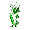 3kvtS S: Starting model for refinement C: citing same article ( |
|---|---|
| Similar structure data | |
| Other databases |
|
-Links
 PDBj
PDBj


- Assembly
| Deposited unit | 
| ||||||||||
|---|---|---|---|---|---|---|---|---|---|---|---|
| 1 |
| ||||||||||
| Unit cell |
| ||||||||||
| Components on special symmetry positions |
|
-Components
| #1: Protein | Mass: 14650.493 Da / Num. of mol.: 5 / Fragment: tetramerization domain (UNP residues 29-147) Source method: isolated from a genetically manipulated source Source: (gene. exp.) Homo sapiens (human) / Gene: KCNB1 / Production host:  #2: Chemical | ChemComp-PEG /   Mass: 106.120 Da / Num. of mol.: 6 / Source method: obtained synthetically / Formula: C4H10O3 Mass: 106.120 Da / Num. of mol.: 6 / Source method: obtained synthetically / Formula: C4H10O3#3: Chemical | ChemComp-ZN /   Mass: 65.409 Da / Num. of mol.: 6 / Source method: obtained synthetically / Formula: Zn / Feature type: SUBJECT OF INVESTIGATION Mass: 65.409 Da / Num. of mol.: 6 / Source method: obtained synthetically / Formula: Zn / Feature type: SUBJECT OF INVESTIGATION#4: Water | ChemComp-HOH / |  Mass: 18.015 Da / Num. of mol.: 13 / Source method: isolated from a natural source / Formula: H2O Mass: 18.015 Da / Num. of mol.: 13 / Source method: isolated from a natural source / Formula: H2OHas ligand of interest | Y | |
|---|
-Experimental details
-Experiment
| Experiment | Method: X-RAY DIFFRACTION / Number of used crystals: 1 |
|---|
- Sample preparation
| Crystal | Density Matthews: 2.35 Å3/Da / Density % sol: 47.69 % |
|---|---|
| Crystal grow | Temperature: 291 K / Method: vapor diffusion, hanging drop / pH: 7.5 Details: 0.2 M magnesium chloride, 15% PEG400, 0.1 M HEPES, pH 7.5 |
-Data collection
| Diffraction | Mean temperature: 100 K / Serial crystal experiment: N | ||||||||||||||||||||||||||||||
|---|---|---|---|---|---|---|---|---|---|---|---|---|---|---|---|---|---|---|---|---|---|---|---|---|---|---|---|---|---|---|---|
| Diffraction source | Source: SYNCHROTRON / Site: ALS  / Beamline: 4.2.2 / Wavelength: 1 Å / Beamline: 4.2.2 / Wavelength: 1 Å | ||||||||||||||||||||||||||||||
| Detector | Type: RDI CMOS_8M / Detector: CMOS / Date: Nov 6, 2020 | ||||||||||||||||||||||||||||||
| Radiation | Protocol: SINGLE WAVELENGTH / Monochromatic (M) / Laue (L): M / Scattering type: x-ray | ||||||||||||||||||||||||||||||
| Radiation wavelength | Wavelength: 1 Å / Relative weight: 1 | ||||||||||||||||||||||||||||||
| Reflection | Resolution: 2.7→48.43 Å / Num. obs: 36640 / % possible obs: 100 % / Redundancy: 7.1 % / Biso Wilson estimate: 56.02 Å2 / CC1/2: 0.998 / Rmerge(I) obs: 0.113 / Rpim(I) all: 0.045 / Rrim(I) all: 0.122 / Net I/σ(I): 17.2 | ||||||||||||||||||||||||||||||
| Reflection shell | Diffraction-ID: 1
|
- Processing
| Software |
| ||||||||||||||||||||||||||||||||||||||||||||||||||||||||||||||||||||||||||||||||||||||||||
|---|---|---|---|---|---|---|---|---|---|---|---|---|---|---|---|---|---|---|---|---|---|---|---|---|---|---|---|---|---|---|---|---|---|---|---|---|---|---|---|---|---|---|---|---|---|---|---|---|---|---|---|---|---|---|---|---|---|---|---|---|---|---|---|---|---|---|---|---|---|---|---|---|---|---|---|---|---|---|---|---|---|---|---|---|---|---|---|---|---|---|---|
| Refinement | Method to determine structure: MOLECULAR REPLACEMENT Starting model: PDB entry 3KVT Resolution: 2.7→45.67 Å / SU ML: 0.39 / Cross valid method: THROUGHOUT / σ(F): 1.33 / Phase error: 27.53 / Stereochemistry target values: ML
| ||||||||||||||||||||||||||||||||||||||||||||||||||||||||||||||||||||||||||||||||||||||||||
| Solvent computation | Shrinkage radii: 0.9 Å / VDW probe radii: 1.11 Å / Solvent model: FLAT BULK SOLVENT MODEL | ||||||||||||||||||||||||||||||||||||||||||||||||||||||||||||||||||||||||||||||||||||||||||
| Displacement parameters | Biso max: 130.11 Å2 / Biso mean: 58.5066 Å2 / Biso min: 29.82 Å2 | ||||||||||||||||||||||||||||||||||||||||||||||||||||||||||||||||||||||||||||||||||||||||||
| Refinement step | Cycle: final / Resolution: 2.7→45.67 Å
| ||||||||||||||||||||||||||||||||||||||||||||||||||||||||||||||||||||||||||||||||||||||||||
| LS refinement shell | Refine-ID: X-RAY DIFFRACTION / Rfactor Rfree error: 0 / Total num. of bins used: 14 / % reflection obs: 100 %
|
