 Movie
Movie Controller

[English] 日本語
 Yorodumi
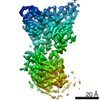
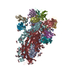
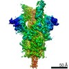
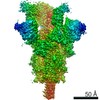
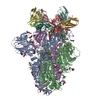
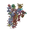
- PDB-7so9: SARS-CoV-2 S B.1.617.2 delta variant + S2M11 + S2L20 Global Refinement -
+ Open data

- Basic information
| Entry | Database: PDB / ID: 7so9 | ||||||
|---|---|---|---|---|---|---|---|
| Title | SARS-CoV-2 S B.1.617.2 delta variant + S2M11 + S2L20 Global Refinement | ||||||
 Components Components |
| ||||||
 Keywords Keywords | VIRAL PROTEIN/IMMUNE SYSTEM / delta / spike / antibody / Structural Genomics / Seattle Structural Genomics Center for Infectious Disease / SSGCID / VIRAL PROTEIN-IMMUNE SYSTEM complex | ||||||
| Function / homology |  Function and homology information Function and homology informationsymbiont-mediated disruption of host tissue / Maturation of spike protein / Translation of Structural Proteins / Virion Assembly and Release / host cell surface / host extracellular space / viral translation / symbiont-mediated-mediated suppression of host tetherin activity / Induction of Cell-Cell Fusion / structural constituent of virion ...symbiont-mediated disruption of host tissue / Maturation of spike protein / Translation of Structural Proteins / Virion Assembly and Release / host cell surface / host extracellular space / viral translation / symbiont-mediated-mediated suppression of host tetherin activity / Induction of Cell-Cell Fusion / structural constituent of virion / entry receptor-mediated virion attachment to host cell / membrane fusion / Attachment and Entry / host cell endoplasmic reticulum-Golgi intermediate compartment membrane / positive regulation of viral entry into host cell / receptor-mediated virion attachment to host cell / host cell surface receptor binding / symbiont-mediated suppression of host innate immune response / receptor ligand activity / endocytosis involved in viral entry into host cell / fusion of virus membrane with host plasma membrane / fusion of virus membrane with host endosome membrane / viral envelope / symbiont entry into host cell / virion attachment to host cell / SARS-CoV-2 activates/modulates innate and adaptive immune responses / host cell plasma membrane / virion membrane / identical protein binding / membrane / plasma membrane Similarity search - Function | ||||||
| Biological species |   Severe acute respiratory syndrome coronavirus 2 Severe acute respiratory syndrome coronavirus 2 Homo sapiens (human) Homo sapiens (human) | ||||||
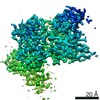
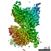
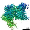
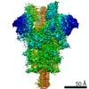
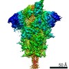
| Method | ELECTRON MICROSCOPY / single particle reconstruction / cryo EM / Resolution: 2.4 Å | ||||||
 Authors Authors | McCallum, M. / Veesler, D. / Seattle Structural Genomics Center for Infectious Disease (SSGCID) | ||||||
| Funding support |  United States, 1items United States, 1items
| ||||||
 Citation Citation | Journal: Science / Year: 2021 Title: Molecular basis of immune evasion by the Delta and Kappa SARS-CoV-2 variants. Authors: Matthew McCallum / Alexandra C Walls / Kaitlin R Sprouse / John E Bowen / Laura E Rosen / Ha V Dang / Anna De Marco / Nicholas Franko / Sasha W Tilles / Jennifer Logue / Marcos C Miranda / ...Authors: Matthew McCallum / Alexandra C Walls / Kaitlin R Sprouse / John E Bowen / Laura E Rosen / Ha V Dang / Anna De Marco / Nicholas Franko / Sasha W Tilles / Jennifer Logue / Marcos C Miranda / Margaret Ahlrichs / Lauren Carter / Gyorgy Snell / Matteo Samuele Pizzuto / Helen Y Chu / Wesley C Van Voorhis / Davide Corti / David Veesler /  Abstract: Severe acute respiratory syndrome coronavirus 2 (SARS-CoV-2) transmission leads to the emergence of variants, including the B.1.617.2 (Delta) variant of concern that is causing a new wave of ...Severe acute respiratory syndrome coronavirus 2 (SARS-CoV-2) transmission leads to the emergence of variants, including the B.1.617.2 (Delta) variant of concern that is causing a new wave of infections and has become globally dominant. We show that these variants dampen the in vitro potency of vaccine-elicited serum neutralizing antibodies and provide a structural framework for describing their immune evasion. Mutations in the B.1.617.1 (Kappa) and Delta spike glycoproteins abrogate recognition by several monoclonal antibodies via alteration of key antigenic sites, including remodeling of the Delta amino-terminal domain. The angiotensin-converting enzyme 2 binding affinities of the Kappa and Delta receptor binding domains are comparable to the Wuhan-Hu-1 isolate, whereas B.1.617.2+ (Delta+) exhibits markedly reduced affinity. | ||||||
| History |
|
- Structure visualization
| Movie |
Movie viewer |
|---|---|
| Structure viewer | Molecule: MolmilJmol/JSmol |
- Downloads & links
-Download
| PDBx/mmCIF format | 7so9.cif.gz | 853.1 KB | Display | PDBx/mmCIF format |
|---|---|---|---|---|
| PDB format | pdb7so9.ent.gz | 678.2 KB | Display | PDB format |
| PDBx/mmJSON format | 7so9.json.gz | Tree view | PDBx/mmJSON format | |
| Others |  Other downloads Other downloads |
-Validation report
| Summary document | 7so9_validation.pdf.gz | 1.3 MB | Display | wwPDB validaton report |
|---|---|---|---|---|
| Full document | 7so9_full_validation.pdf.gz | 1.3 MB | Display | |
| Data in XML | 7so9_validation.xml.gz | 115.8 KB | Display | |
| Data in CIF | 7so9_validation.cif.gz | 181.2 KB | Display | |
| Arichive directory | https://data.pdbj.org/pub/pdb/validation_reports/so/7so9ftp://data.pdbj.org/pub/pdb/validation_reports/so/7so9 | HTTPS FTP |
-Related structure data
| Related structure data | 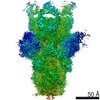 25263MC 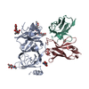 7soaC 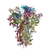 7sobC  7socC 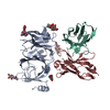 7sodC  7soeC 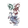 7sofC C: citing same article ( M: map data used to model this data |
|---|---|
| Similar structure data |


-Links
 PDBj
PDBj


- Assembly
| Deposited unit | 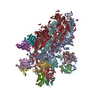
|
|---|---|
| 1 |
|
-Components
-Antibody , 4 types, 12 molecules DGLEHMBINCJO
| #2: Antibody | Mass: 11208.458 Da / Num. of mol.: 3 Source method: isolated from a genetically manipulated source Source: (gene. exp.) Homo sapiens (human) / Production host: Homo sapiens (human)#3: Antibody | Mass: 13651.220 Da / Num. of mol.: 3 Source method: isolated from a genetically manipulated source Source: (gene. exp.) Homo sapiens (human) / Production host: Homo sapiens (human)#4: Antibody | Mass: 11829.141 Da / Num. of mol.: 3 Source method: isolated from a genetically manipulated source Source: (gene. exp.) Homo sapiens (human) / Production host: Homo sapiens (human)#5: Antibody | Mass: 13453.958 Da / Num. of mol.: 3 Source method: isolated from a genetically manipulated source Source: (gene. exp.) Homo sapiens (human) / Production host: Homo sapiens (human) |
|---|
-Protein / Non-polymers , 2 types, 357 molecules AFK
| #1: Protein | Mass: 141522.891 Da / Num. of mol.: 3 Source method: isolated from a genetically manipulated source Source: (gene. exp.) Severe acute respiratory syndrome coronavirus 2Gene: S, 2 / Production host: Homo sapiens (human) / References: UniProt: P0DTC2#8: Water | ChemComp-HOH / | Mass: 18.015 Da / Num. of mol.: 354 / Source method: isolated from a natural source / Formula: H2O |
|---|
-Sugars , 2 types, 48 molecules 
| #6: Polysaccharide | | #7: Sugar | ChemComp-NAG /  Type: D-saccharide, beta linking / Mass: 221.208 Da / Num. of mol.: 45 / Source method: obtained synthetically / Formula: C8H15NO6 Type: D-saccharide, beta linking / Mass: 221.208 Da / Num. of mol.: 45 / Source method: obtained synthetically / Formula: C8H15NO6 |
|---|
-Details
| Has ligand of interest | N |
|---|---|
| Has protein modification | Y |
-Experimental details
-Experiment
| Experiment | Method: ELECTRON MICROSCOPY |
|---|---|
| EM experiment | Aggregation state: PARTICLE / 3D reconstruction method: single particle reconstruction |
- Sample preparation
| Component | Name: SARS-CoV-2 S B.1.617.2 delta variant + S2M11 Fab + S2L20 Fab Type: COMPLEX / Entity ID: #1-#5 / Source: MULTIPLE SOURCES | ||||||||||||
|---|---|---|---|---|---|---|---|---|---|---|---|---|---|
| Source (natural) |
| ||||||||||||
| Source (recombinant) | Organism: Homo sapiens (human) | ||||||||||||
| Buffer solution | pH: 8 | ||||||||||||
| Specimen | Embedding applied: NO / Shadowing applied: NO / Staining applied: NO / Vitrification applied: YES | ||||||||||||
| Vitrification | Cryogen name: ETHANE |
- Electron microscopy imaging
| Experimental equipment |  Model: Titan Krios / Image courtesy: FEI Company |
|---|---|
| Microscopy | Model: FEI TITAN KRIOS |
| Electron gun | Electron source:  FIELD EMISSION GUN / Accelerating voltage: 300 kV / Illumination mode: FLOOD BEAM FIELD EMISSION GUN / Accelerating voltage: 300 kV / Illumination mode: FLOOD BEAM |
| Electron lens | Mode: BRIGHT FIELD |
| Image recording | Electron dose: 63 e/Å2 / Film or detector model: GATAN K3 (6k x 4k) |
- Processing
| CTF correction | Type: PHASE FLIPPING AND AMPLITUDE CORRECTION |
|---|---|
| 3D reconstruction | Resolution: 2.4 Å / Resolution method: FSC 0.143 CUT-OFF / Num. of particles: 543668 / Symmetry type: POINT |
