 Movie
Movie Controller

[English] 日本語
 Yorodumi
- PDB-7so8: Crystal structure of Glutathione S-Transferase from Shrimp Litope... -
+ Open data

- Basic information
| Entry | Database: PDB / ID: 7so8 | ||||||
|---|---|---|---|---|---|---|---|
| Title | Crystal structure of Glutathione S-Transferase from Shrimp Litopenaeus vannamei in complex with silver ions and a molecules of Glutathione binding in G-site and H-site | ||||||
 Components Components | Glutathione transferase | ||||||
 Keywords Keywords | TRANSFERASE / GST / GSH binding in G-site | ||||||
| Function / homology |  Function and homology information Function and homology informationglutathione transferase / glutathione transferase activity / glutathione metabolic process Similarity search - Function | ||||||
| Biological species |  Penaeus vannamei (Pacific white shrimp) Penaeus vannamei (Pacific white shrimp) | ||||||
| Method |  X-RAY DIFFRACTION / SYNCHROTRON / MOLECULAR REPLACEMENT / molecular replacement / Resolution: 2.2 Å X-RAY DIFFRACTION / SYNCHROTRON / MOLECULAR REPLACEMENT / molecular replacement / Resolution: 2.2 Å | ||||||
 Authors Authors | Escudero-Garcia, A. / Rudino-Pinera, E. / Miranda-Blancas, R. | ||||||
| Funding support | 1items
| ||||||
 Citation Citation | Journal: To Be Published Title: Inhibition of GST class Mu of the shrimp Litopenaeus vannamei by binding silver ions in H-site. Authors: Escudero-Garcia, A. / Rudino-Pinera, E. | ||||||
| History |
|
- Structure visualization
| Structure viewer | Molecule: MolmilJmol/JSmol |
|---|
- Downloads & links
-Download
| PDBx/mmCIF format | 7so8.cif.gz | 388.4 KB | Display | PDBx/mmCIF format |
|---|---|---|---|---|
| PDB format | pdb7so8.ent.gz | 317.4 KB | Display | PDB format |
| PDBx/mmJSON format | 7so8.json.gz | Tree view | PDBx/mmJSON format | |
| Others |  Other downloads Other downloads |
-Validation report
| Summary document | 7so8_validation.pdf.gz | 3.6 MB | Display | wwPDB validaton report |
|---|---|---|---|---|
| Full document | 7so8_full_validation.pdf.gz | 3.5 MB | Display | |
| Data in XML | 7so8_validation.xml.gz | 78.6 KB | Display | |
| Data in CIF | 7so8_validation.cif.gz | 109 KB | Display | |
| Arichive directory | https://data.pdbj.org/pub/pdb/validation_reports/so/7so8ftp://data.pdbj.org/pub/pdb/validation_reports/so/7so8 | HTTPS FTP |
-Related structure data
| Related structure data | 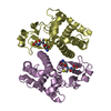 5an1S S: Starting model for refinement |
|---|---|
| Similar structure data |
-Links
 PDBj
PDBj

- Assembly
| Deposited unit | 
| ||||||||
|---|---|---|---|---|---|---|---|---|---|
| 1 | 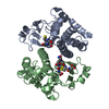
| ||||||||
| 2 | 
| ||||||||
| 3 | 
| ||||||||
| 4 | 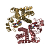
| ||||||||
| Unit cell |
|
-Components
| #1: Protein | Mass: 25552.455 Da / Num. of mol.: 8 Source method: isolated from a genetically manipulated source Source: (gene. exp.) Penaeus vannamei (Pacific white shrimp)Production host:  #2: Chemical | ChemComp-GSH /   Mass: 307.323 Da / Num. of mol.: 10 / Source method: obtained synthetically / Formula: C10H17N3O6S / Feature type: SUBJECT OF INVESTIGATION Mass: 307.323 Da / Num. of mol.: 10 / Source method: obtained synthetically / Formula: C10H17N3O6S / Feature type: SUBJECT OF INVESTIGATION#3: Chemical | ChemComp-1PE / |   Mass: 238.278 Da / Num. of mol.: 1 / Source method: obtained synthetically / Formula: C10H22O6 / Comment: precipitant*YM Mass: 238.278 Da / Num. of mol.: 1 / Source method: obtained synthetically / Formula: C10H22O6 / Comment: precipitant*YM#4: Chemical |   Mass: 107.868 Da / Num. of mol.: 3 / Source method: obtained synthetically / Formula: Ag / Feature type: SUBJECT OF INVESTIGATION Mass: 107.868 Da / Num. of mol.: 3 / Source method: obtained synthetically / Formula: Ag / Feature type: SUBJECT OF INVESTIGATION#5: Water | ChemComp-HOH / |  Mass: 18.015 Da / Num. of mol.: 1162 / Source method: isolated from a natural source / Formula: H2O Mass: 18.015 Da / Num. of mol.: 1162 / Source method: isolated from a natural source / Formula: H2OHas ligand of interest | Y | |
|---|
-Experimental details
-Experiment
| Experiment | Method: X-RAY DIFFRACTION / Number of used crystals: 1 |
|---|
- Sample preparation
| Crystal | Density Matthews: 2.21 Å3/Da / Density % sol: 44.4 % |
|---|---|
| Crystal grow | Temperature: 277.15 K / Method: microbatch / pH: 6.5 Details: 100 mM ammonium sulphate, 100 Bis-Tris pH 6.5, 30% PEG 3350, 300 uM silver nitrate PH range: 6.5-8 |
-Data collection
| Diffraction | Mean temperature: 100 K / Serial crystal experiment: N | ||||||||||||||||||||||||||||||||||||||||
|---|---|---|---|---|---|---|---|---|---|---|---|---|---|---|---|---|---|---|---|---|---|---|---|---|---|---|---|---|---|---|---|---|---|---|---|---|---|---|---|---|---|
| Diffraction source | Source: SYNCHROTRON / Site: APS  / Beamline: 19-BM / Wavelength: 0.9793 Å / Beamline: 19-BM / Wavelength: 0.9793 Å | ||||||||||||||||||||||||||||||||||||||||
| Detector | Type: ADSC QUANTUM 210r / Detector: CCD / Date: Jun 12, 2019 / Details: Sagitally focusing 2nd Crystal | ||||||||||||||||||||||||||||||||||||||||
| Radiation | Protocol: SINGLE WAVELENGTH / Monochromatic (M) / Laue (L): M / Scattering type: x-ray | ||||||||||||||||||||||||||||||||||||||||
| Radiation wavelength | Wavelength: 0.9793 Å / Relative weight: 1 | ||||||||||||||||||||||||||||||||||||||||
| Reflection | Resolution: 2.2→56.37 Å / Num. obs: 88718 / % possible obs: 98.15 % / Redundancy: 3.104 % / Biso Wilson estimate: 13.76 Å2 / CC1/2: 0.627 / Rmerge(I) obs: 0.6179 / Rpim(I) all: 0.4226 / Rrim(I) all: 0.7511 / Net I/av σ(I): 1.98 / Net I/σ(I): 6.6 | ||||||||||||||||||||||||||||||||||||||||
| Reflection shell | Diffraction-ID: 1 / Redundancy: 3.1 %
|
-Phasing
| Phasing | Method: molecular replacement |
|---|
- Processing
| Software |
| |||||||||||||||||||||||||||||||||||||||||||||||||||||||||||||||||||||||||||||||||||||||||||||||||||||||||||||||||||||||||||||||||||||||||||||||||||||||||||||||||||||||||||||||||||||||||||||||||||||||||||||||||||||||||
|---|---|---|---|---|---|---|---|---|---|---|---|---|---|---|---|---|---|---|---|---|---|---|---|---|---|---|---|---|---|---|---|---|---|---|---|---|---|---|---|---|---|---|---|---|---|---|---|---|---|---|---|---|---|---|---|---|---|---|---|---|---|---|---|---|---|---|---|---|---|---|---|---|---|---|---|---|---|---|---|---|---|---|---|---|---|---|---|---|---|---|---|---|---|---|---|---|---|---|---|---|---|---|---|---|---|---|---|---|---|---|---|---|---|---|---|---|---|---|---|---|---|---|---|---|---|---|---|---|---|---|---|---|---|---|---|---|---|---|---|---|---|---|---|---|---|---|---|---|---|---|---|---|---|---|---|---|---|---|---|---|---|---|---|---|---|---|---|---|---|---|---|---|---|---|---|---|---|---|---|---|---|---|---|---|---|---|---|---|---|---|---|---|---|---|---|---|---|---|---|---|---|---|---|---|---|---|---|---|---|---|---|---|---|---|---|---|---|---|
| Refinement | Method to determine structure: MOLECULAR REPLACEMENT Starting model: 5AN1 Resolution: 2.2→56.37 Å / SU ML: 0.32 / Cross valid method: THROUGHOUT / σ(F): 1.98 / Phase error: 25.86 / Stereochemistry target values: ML
| |||||||||||||||||||||||||||||||||||||||||||||||||||||||||||||||||||||||||||||||||||||||||||||||||||||||||||||||||||||||||||||||||||||||||||||||||||||||||||||||||||||||||||||||||||||||||||||||||||||||||||||||||||||||||
| Solvent computation | Shrinkage radii: 0.9 Å / VDW probe radii: 1.11 Å / Solvent model: FLAT BULK SOLVENT MODEL | |||||||||||||||||||||||||||||||||||||||||||||||||||||||||||||||||||||||||||||||||||||||||||||||||||||||||||||||||||||||||||||||||||||||||||||||||||||||||||||||||||||||||||||||||||||||||||||||||||||||||||||||||||||||||
| Displacement parameters | Biso max: 134.29 Å2 / Biso mean: 16.3226 Å2 / Biso min: 1.6 Å2 | |||||||||||||||||||||||||||||||||||||||||||||||||||||||||||||||||||||||||||||||||||||||||||||||||||||||||||||||||||||||||||||||||||||||||||||||||||||||||||||||||||||||||||||||||||||||||||||||||||||||||||||||||||||||||
| Refinement step | Cycle: final / Resolution: 2.2→56.37 Å
| |||||||||||||||||||||||||||||||||||||||||||||||||||||||||||||||||||||||||||||||||||||||||||||||||||||||||||||||||||||||||||||||||||||||||||||||||||||||||||||||||||||||||||||||||||||||||||||||||||||||||||||||||||||||||
| LS refinement shell | Refine-ID: X-RAY DIFFRACTION / Rfactor Rfree error: 0 / Total num. of bins used: 30
|
