 Movie
Movie Controller

[English] 日本語
 Yorodumi
- PDB-7snu: Crystal structure of ShHTL7 from Striga hermonthica in complex wi... -
+ Open data

- Basic information
| Entry | Database: PDB / ID: 7snu | |||||||||||||||||||||
|---|---|---|---|---|---|---|---|---|---|---|---|---|---|---|---|---|---|---|---|---|---|---|
| Title | Crystal structure of ShHTL7 from Striga hermonthica in complex with strigolactone antagonist RG6 | |||||||||||||||||||||
 Components Components | Hyposensitive to light 7 | |||||||||||||||||||||
 Keywords Keywords | HYDROLASE / Parasite / Strigolactone / Receptor / Antagonist | |||||||||||||||||||||
| Function / homology | Alpha/beta hydrolase family / Alpha/beta hydrolase fold-1 / Alpha/Beta hydrolase fold / hydrolase activity / metal ion binding / ACETATE ION / Chem-HFJ / IMIDAZOLE / Hyposensitive to light 7 Function and homology information Function and homology information | |||||||||||||||||||||
| Biological species |  Striga hermonthica (purple witchweed) Striga hermonthica (purple witchweed) | |||||||||||||||||||||
| Method |  X-RAY DIFFRACTION / SYNCHROTRON / MOLECULAR REPLACEMENT / Resolution: 1.46 Å X-RAY DIFFRACTION / SYNCHROTRON / MOLECULAR REPLACEMENT / Resolution: 1.46 Å | |||||||||||||||||||||
 Authors Authors | Arellano-Saab, A. / Stogios, P.J. / Skarina, T. / Yim, V. / Savchenko, A. / McCourt, P. | |||||||||||||||||||||
| Funding support | 6items
| |||||||||||||||||||||
 Citation Citation | Journal: J.Biol.Chem. / Year: 2022 Title: A novel strigolactone receptor antagonist provides insights into the structural inhibition, conditioning, and germination of the crop parasite Striga. Authors: Arellano-Saab, A. / McErlean, C.S.P. / Lumba, S. / Savchenko, A. / Stogios, P.J. / McCourt, P. | |||||||||||||||||||||
| History |
|
- Structure visualization
| Structure viewer | Molecule: MolmilJmol/JSmol |
|---|
- Downloads & links
-Download
| PDBx/mmCIF format | 7snu.cif.gz | 161.8 KB | Display | PDBx/mmCIF format |
|---|---|---|---|---|
| PDB format | pdb7snu.ent.gz | 103.3 KB | Display | PDB format |
| PDBx/mmJSON format | 7snu.json.gz | Tree view | PDBx/mmJSON format | |
| Others |  Other downloads Other downloads |
-Validation report
| Arichive directory | https://data.pdbj.org/pub/pdb/validation_reports/sn/7snuftp://data.pdbj.org/pub/pdb/validation_reports/sn/7snu | HTTPS FTP |
|---|
-Related structure data
| Related structure data |  5cbkS S: Starting model for refinement |
|---|---|
| Similar structure data |
-Links
 PDBj
PDBj


- Assembly
| Deposited unit | 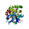
| ||||||||||||
|---|---|---|---|---|---|---|---|---|---|---|---|---|---|
| 1 |
| ||||||||||||
| Unit cell |
|
-Components
-Protein , 1 types, 1 molecules A
| #1: Protein | Mass: 30255.768 Da / Num. of mol.: 1 Source method: isolated from a genetically manipulated source Source: (gene. exp.) Striga hermonthica (purple witchweed) / Plasmid: pMCSG53 / Production host:  |
|---|
-Non-polymers , 6 types, 484 molecules 


| #2: Chemical |  Mass: 92.094 Da / Num. of mol.: 2 / Source method: obtained synthetically / Formula: C3H8O3 Mass: 92.094 Da / Num. of mol.: 2 / Source method: obtained synthetically / Formula: C3H8O3#3: Chemical | ChemComp-ACT /  Mass: 59.044 Da / Num. of mol.: 5 / Source method: obtained synthetically / Formula: C2H3O2 Mass: 59.044 Da / Num. of mol.: 5 / Source method: obtained synthetically / Formula: C2H3O2#4: Chemical | ChemComp-MG / |  Mass: 24.305 Da / Num. of mol.: 1 / Source method: obtained synthetically / Formula: Mg Mass: 24.305 Da / Num. of mol.: 1 / Source method: obtained synthetically / Formula: Mg#5: Chemical | ChemComp-IMD / |  Mass: 69.085 Da / Num. of mol.: 1 / Source method: obtained synthetically / Formula: C3H5N2 Mass: 69.085 Da / Num. of mol.: 1 / Source method: obtained synthetically / Formula: C3H5N2#6: Chemical | ChemComp-HFJ / |  Mass: 410.549 Da / Num. of mol.: 1 / Source method: obtained synthetically / Formula: C25H34N2O3 / Feature type: SUBJECT OF INVESTIGATION Mass: 410.549 Da / Num. of mol.: 1 / Source method: obtained synthetically / Formula: C25H34N2O3 / Feature type: SUBJECT OF INVESTIGATION#7: Water | ChemComp-HOH / | Mass: 18.015 Da / Num. of mol.: 474 / Source method: isolated from a natural source / Formula: H2O |
|---|
-Details
| Has ligand of interest | Y |
|---|
-Experimental details
-Experiment
| Experiment | Method: X-RAY DIFFRACTION / Number of used crystals: 1 |
|---|
- Sample preparation
| Crystal | Density Matthews: 3.07 Å3/Da / Density % sol: 59.95 % |
|---|---|
| Crystal grow | Temperature: 298 K / Method: vapor diffusion, hanging drop / pH: 7 Details: 1 M sodium acetate, 0.1 M imidazole pH 7.0, 5% glycerol |
-Data collection
| Diffraction | Mean temperature: 100 K / Serial crystal experiment: N |
|---|---|
| Diffraction source | Source: SYNCHROTRON / Site: APS  / Beamline: 21-ID-D / Wavelength: 0.97913 Å / Beamline: 21-ID-D / Wavelength: 0.97913 Å |
| Detector | Type: DECTRIS EIGER X 9M / Detector: PIXEL / Date: Apr 11, 2018 |
| Radiation | Monochromator: Si(111) / Protocol: SINGLE WAVELENGTH / Monochromatic (M) / Laue (L): M / Scattering type: x-ray |
| Radiation wavelength | Wavelength: 0.97913 Å / Relative weight: 1 |
| Reflection | Resolution: 1.46→30 Å / Num. obs: 62389 / % possible obs: 98.4 % / Redundancy: 6.9 % / Biso Wilson estimate: 12.94 Å2 / Rmerge(I) obs: 0.059 / Rpim(I) all: 0.023 / Net I/σ(I): 28.67 |
| Reflection shell | Resolution: 1.46→1.49 Å / Redundancy: 4.2 % / Rmerge(I) obs: 0.46 / Mean I/σ(I) obs: 2.4 / Num. unique obs: 2550 / CC1/2: 0.841 / Rpim(I) all: 0.233 / % possible all: 80.8 |
- Processing
| Software |
| |||||||||||||||||||||||||||||||||||||||||||||||||||||||||||||||||||||||||||||||||||||||||||||||||||||||||
|---|---|---|---|---|---|---|---|---|---|---|---|---|---|---|---|---|---|---|---|---|---|---|---|---|---|---|---|---|---|---|---|---|---|---|---|---|---|---|---|---|---|---|---|---|---|---|---|---|---|---|---|---|---|---|---|---|---|---|---|---|---|---|---|---|---|---|---|---|---|---|---|---|---|---|---|---|---|---|---|---|---|---|---|---|---|---|---|---|---|---|---|---|---|---|---|---|---|---|---|---|---|---|---|---|---|---|
| Refinement | Method to determine structure: MOLECULAR REPLACEMENT Starting model: PDB entry 5CBK Resolution: 1.46→28.68 Å / SU ML: 0.1155 / Cross valid method: FREE R-VALUE / σ(F): 0 / Phase error: 15.2135 Stereochemistry target values: GeoStd + Monomer Library + CDL v1.2
| |||||||||||||||||||||||||||||||||||||||||||||||||||||||||||||||||||||||||||||||||||||||||||||||||||||||||
| Solvent computation | Shrinkage radii: 0.9 Å / VDW probe radii: 1.11 Å / Solvent model: FLAT BULK SOLVENT MODEL | |||||||||||||||||||||||||||||||||||||||||||||||||||||||||||||||||||||||||||||||||||||||||||||||||||||||||
| Displacement parameters | Biso mean: 21.09 Å2 | |||||||||||||||||||||||||||||||||||||||||||||||||||||||||||||||||||||||||||||||||||||||||||||||||||||||||
| Refinement step | Cycle: LAST / Resolution: 1.46→28.68 Å
| |||||||||||||||||||||||||||||||||||||||||||||||||||||||||||||||||||||||||||||||||||||||||||||||||||||||||
| Refine LS restraints |
| |||||||||||||||||||||||||||||||||||||||||||||||||||||||||||||||||||||||||||||||||||||||||||||||||||||||||
| LS refinement shell |
|
