+ Open data
- Basic information
| Entry | Database: PDB / ID: 7sjo | ||||||||||||||||||||||||||||||||||||||||||||||||||||||||||||||||||||||||
|---|---|---|---|---|---|---|---|---|---|---|---|---|---|---|---|---|---|---|---|---|---|---|---|---|---|---|---|---|---|---|---|---|---|---|---|---|---|---|---|---|---|---|---|---|---|---|---|---|---|---|---|---|---|---|---|---|---|---|---|---|---|---|---|---|---|---|---|---|---|---|---|---|---|
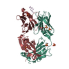
| Title | HtrA1S328A:Fab15H6.v4 complex | ||||||||||||||||||||||||||||||||||||||||||||||||||||||||||||||||||||||||
 Components Components |
| ||||||||||||||||||||||||||||||||||||||||||||||||||||||||||||||||||||||||
 Keywords Keywords | HYDROLASE/Immune System / HtrA1 / allosteric inhibition / Age-related macular degeneration / Geographic Atrophy / Antibody complex / HYDROLASE / HYDROLASE-Immune System complex | ||||||||||||||||||||||||||||||||||||||||||||||||||||||||||||||||||||||||
| Function / homology |  Function and homology information Function and homology informationchorionic trophoblast cell differentiation / programmed cell death / growth factor binding / Hydrolases; Acting on peptide bonds (peptidases); Serine endopeptidases / negative regulation of BMP signaling pathway / Degradation of the extracellular matrix / serine-type peptidase activity / molecular function activator activity / placenta development / negative regulation of transforming growth factor beta receptor signaling pathway ...chorionic trophoblast cell differentiation / programmed cell death / growth factor binding / Hydrolases; Acting on peptide bonds (peptidases); Serine endopeptidases / negative regulation of BMP signaling pathway / Degradation of the extracellular matrix / serine-type peptidase activity / molecular function activator activity / placenta development / negative regulation of transforming growth factor beta receptor signaling pathway / : / positive regulation of apoptotic process / serine-type endopeptidase activity / proteolysis / extracellular space / extracellular exosome / extracellular region / identical protein binding / plasma membrane / cytosol Similarity search - Function | ||||||||||||||||||||||||||||||||||||||||||||||||||||||||||||||||||||||||
| Biological species |  Homo sapiens (human) Homo sapiens (human) | ||||||||||||||||||||||||||||||||||||||||||||||||||||||||||||||||||||||||
| Method | ELECTRON MICROSCOPY / single particle reconstruction / cryo EM / Resolution: 3.3 Å | ||||||||||||||||||||||||||||||||||||||||||||||||||||||||||||||||||||||||
 Authors Authors | Gerhardy, S. / Green, E. / Estevez, A. / Arthur, C.P. / Ultsch, M. / Rohou, A. / Kirchhofer, D. | ||||||||||||||||||||||||||||||||||||||||||||||||||||||||||||||||||||||||
| Funding support |  United States, 1items United States, 1items
| ||||||||||||||||||||||||||||||||||||||||||||||||||||||||||||||||||||||||
 Citation Citation |  Journal: Nat Commun / Year: 2022 Journal: Nat Commun / Year: 2022Title: Allosteric inhibition of HTRA1 activity by a conformational lock mechanism to treat age-related macular degeneration. Authors: Stefan Gerhardy / Mark Ultsch / Wanjian Tang / Evan Green / Jeffrey K Holden / Wei Li / Alberto Estevez / Chris Arthur / Irene Tom / Alexis Rohou / Daniel Kirchhofer /  Abstract: The trimeric serine protease HTRA1 is a genetic risk factor associated with geographic atrophy (GA), a currently untreatable form of age-related macular degeneration. Here, we describe the allosteric ...The trimeric serine protease HTRA1 is a genetic risk factor associated with geographic atrophy (GA), a currently untreatable form of age-related macular degeneration. Here, we describe the allosteric inhibition mechanism of HTRA1 by a clinical Fab fragment, currently being evaluated for GA treatment. Using cryo-EM, X-ray crystallography and biochemical assays we identify the exposed LoopA of HTRA1 as the sole Fab epitope, which is approximately 30 Å away from the active site. The cryo-EM structure of the HTRA1:Fab complex in combination with molecular dynamics simulations revealed that Fab binding to LoopA locks HTRA1 in a non-competent conformational state, incapable of supporting catalysis. Moreover, grafting the HTRA1-LoopA epitope onto HTRA2 and HTRA3 transferred the allosteric inhibition mechanism. This suggests a conserved conformational lock mechanism across the HTRA family and a critical role of LoopA for catalysis, which was supported by the reduced activity of HTRA1-3 upon LoopA deletion or perturbation. This study reveals the long-range inhibition mechanism of the clinical Fab and identifies an essential function of the exposed LoopA for activity of HTRA family proteases. | ||||||||||||||||||||||||||||||||||||||||||||||||||||||||||||||||||||||||
| History |
|
- Structure visualization
| Structure viewer | Molecule:  Molmil Molmil Jmol/JSmol Jmol/JSmol |
|---|
- Downloads & links
- Download
| PDBx/mmCIF format |  7sjo.cif.gz 7sjo.cif.gz | 324.9 KB | Display |  PDBx/mmCIF format PDBx/mmCIF format |
|---|---|---|---|---|
| PDB format |  pdb7sjo.ent.gz pdb7sjo.ent.gz | 261.7 KB | Display |  PDB format PDB format |
| PDBx/mmJSON format |  7sjo.json.gz 7sjo.json.gz | Tree view |  PDBx/mmJSON format PDBx/mmJSON format | |
| Others |  Other downloads Other downloads |
-Validation report
| Summary document |  7sjo_validation.pdf.gz 7sjo_validation.pdf.gz | 1.2 MB | Display |  wwPDB validaton report wwPDB validaton report |
|---|---|---|---|---|
| Full document |  7sjo_full_validation.pdf.gz 7sjo_full_validation.pdf.gz | 1.2 MB | Display | |
| Data in XML |  7sjo_validation.xml.gz 7sjo_validation.xml.gz | 60.3 KB | Display | |
| Data in CIF |  7sjo_validation.cif.gz 7sjo_validation.cif.gz | 88.6 KB | Display | |
| Arichive directory |  https://data.pdbj.org/pub/pdb/validation_reports/sj/7sjo https://data.pdbj.org/pub/pdb/validation_reports/sj/7sjo ftp://data.pdbj.org/pub/pdb/validation_reports/sj/7sjo ftp://data.pdbj.org/pub/pdb/validation_reports/sj/7sjo | HTTPS FTP |
-Related structure data
| Related structure data |  25163MC  7sjmC  7sjnC  7sjpC M: map data used to model this data C: citing same article ( |
|---|---|
| Similar structure data | Similarity search - Function & homology  F&H Search F&H Search |
- Links
Links
- Assembly
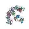
| Deposited unit | 
|
|---|---|
| 1 |
|
- Components
| #1: Protein | Mass: 25714.385 Da / Num. of mol.: 3 / Mutation: S328A Source method: isolated from a genetically manipulated source Source: (gene. exp.)  Homo sapiens (human) / Gene: HTRA1, HTRA, PRSS11 / Variant: S328A / Production host: Homo sapiens (human) / Gene: HTRA1, HTRA, PRSS11 / Variant: S328A / Production host:  References: UniProt: Q92743, Hydrolases; Acting on peptide bonds (peptidases); Serine endopeptidases #2: Antibody | Mass: 26768.996 Da / Num. of mol.: 3 Source method: isolated from a genetically manipulated source Source: (gene. exp.)  Homo sapiens (human) / Production host: Homo sapiens (human) / Production host:  #3: Antibody | Mass: 25717.730 Da / Num. of mol.: 3 Source method: isolated from a genetically manipulated source Source: (gene. exp.)  Homo sapiens (human) / Production host: Homo sapiens (human) / Production host:  Has protein modification | Y | |
|---|
-Experimental details
-Experiment
| Experiment | Method: ELECTRON MICROSCOPY |
|---|---|
| EM experiment | Aggregation state: PARTICLE / 3D reconstruction method: single particle reconstruction |
- Sample preparation
| Component | Name: HtrA1PD/SA bound by Fab15H6.v4 at LoopA epitope / Type: COMPLEX / Details: clinical Fab fragment Fab15H6.v4 / Entity ID: all / Source: RECOMBINANT | ||||||||||||||||||||
|---|---|---|---|---|---|---|---|---|---|---|---|---|---|---|---|---|---|---|---|---|---|
| Molecular weight | Value: 0.22 MDa / Experimental value: NO | ||||||||||||||||||||
| Source (natural) | Organism:  Homo sapiens (human) Homo sapiens (human) | ||||||||||||||||||||
| Source (recombinant) | Organism:  | ||||||||||||||||||||
| Buffer solution | pH: 7.2 | ||||||||||||||||||||
| Buffer component |
| ||||||||||||||||||||
| Specimen | Conc.: 4 mg/ml / Embedding applied: NO / Shadowing applied: NO / Staining applied: NO / Vitrification applied: YES / Details: HtrA1PD/SA:Fab15H6.v4 complex | ||||||||||||||||||||
| Specimen support | Details: SAM: Grids were incubated in 4 mM monothiolalkane(C11)PEG6-OH (11-mercaptoundecyl) hexaethylenglycol (SPT-0011P6, SensoPath Technologies, Inc., Bozeman, MT) for 24h and rinsed in EtOH before sample application Grid material: GOLD / Grid mesh size: 400 divisions/in. / Grid type: Quantifoil R0.6/1 | ||||||||||||||||||||
| Vitrification | Instrument: LEICA EM GP / Cryogen name: ETHANE / Humidity: 100 % / Chamber temperature: 277 K / Details: 3.5s blot time |
- Electron microscopy imaging
| Experimental equipment |  Model: Titan Krios / Image courtesy: FEI Company |
|---|---|
| Microscopy | Model: FEI TITAN KRIOS |
| Electron gun | Electron source:  FIELD EMISSION GUN / Accelerating voltage: 300 kV / Illumination mode: FLOOD BEAM FIELD EMISSION GUN / Accelerating voltage: 300 kV / Illumination mode: FLOOD BEAM |
| Electron lens | Mode: BRIGHT FIELD / Nominal magnification: 105000 X / Nominal defocus max: 1500 nm / Nominal defocus min: 500 nm / Cs: 2.7 mm / C2 aperture diameter: 50 µm |
| Specimen holder | Cryogen: NITROGEN / Specimen holder model: FEI TITAN KRIOS AUTOGRID HOLDER |
| Image recording | Average exposure time: 3 sec. / Electron dose: 65 e/Å2 / Film or detector model: GATAN K3 BIOQUANTUM (6k x 4k) / Num. of grids imaged: 1 / Num. of real images: 7336 |
- Processing
| Software | Name: PHENIX / Version: 1.19.1-4122_final: / Classification: refinement | ||||||||||||||||||||||||
|---|---|---|---|---|---|---|---|---|---|---|---|---|---|---|---|---|---|---|---|---|---|---|---|---|---|
| EM software |
| ||||||||||||||||||||||||
| CTF correction | Type: PHASE FLIPPING AND AMPLITUDE CORRECTION | ||||||||||||||||||||||||
| Particle selection | Num. of particles selected: 505669 | ||||||||||||||||||||||||
| Symmetry | Point symmetry: C1 (asymmetric) | ||||||||||||||||||||||||
| 3D reconstruction | Resolution: 3.3 Å / Resolution method: FSC 0.143 CUT-OFF / Num. of particles: 219676 / Num. of class averages: 1 / Symmetry type: POINT | ||||||||||||||||||||||||
| Atomic model building | Protocol: RIGID BODY FIT | ||||||||||||||||||||||||
| Atomic model building | PDB-ID: 3TJN Pdb chain-ID: A / Accession code: 3TJN / Source name: PDB / Type: experimental model | ||||||||||||||||||||||||
| Refine LS restraints |
|
 Movie
Movie Controller


 PDBj
PDBj


