[English] 日本語
 Yorodumi
- PDB-7sit: Crystal structure of Voltage gated potassium ion channel, Kv 1.2 ... -
+ Open data
- Basic information
| Entry | Database: PDB / ID: 7sit | ||||||
|---|---|---|---|---|---|---|---|
| Title | Crystal structure of Voltage gated potassium ion channel, Kv 1.2 chimera-3m | ||||||
 Components Components |
| ||||||
 Keywords Keywords | TRANSPORT PROTEIN / OXIDOREDUCTASE / Ion channel / C-type inactivation / Voltage gated Potassium ion channel / Chimera | ||||||
| Function / homology |  Function and homology information Function and homology informationpinceau fiber / methylglyoxal reductase (NADPH) (acetol producing) activity / Voltage gated Potassium channels / potassium channel complex / regulation of protein localization to cell surface / : / axon initial segment / juxtaparanode region of axon / Oxidoreductases; Acting on the CH-OH group of donors; With NAD+ or NADP+ as acceptor / myoblast differentiation ...pinceau fiber / methylglyoxal reductase (NADPH) (acetol producing) activity / Voltage gated Potassium channels / potassium channel complex / regulation of protein localization to cell surface / : / axon initial segment / juxtaparanode region of axon / Oxidoreductases; Acting on the CH-OH group of donors; With NAD+ or NADP+ as acceptor / myoblast differentiation / regulation of potassium ion transmembrane transport / Neutrophil degranulation / neuromuscular process / voltage-gated potassium channel activity / potassium channel regulator activity / hematopoietic progenitor cell differentiation / axon terminus / voltage-gated potassium channel complex / postsynaptic density membrane / cytoplasmic side of plasma membrane / transmembrane transporter binding / cytoskeleton / neuron projection / postsynaptic density / axon / protein-containing complex binding / glutamatergic synapse / membrane / cytosol Similarity search - Function | ||||||
| Biological species |  | ||||||
| Method |  X-RAY DIFFRACTION / X-RAY DIFFRACTION /  SYNCHROTRON / SYNCHROTRON /  MOLECULAR REPLACEMENT / MOLECULAR REPLACEMENT /  molecular replacement / Resolution: 3.32 Å molecular replacement / Resolution: 3.32 Å | ||||||
 Authors Authors | Reddi, R. / Matulef, K. / Riederer, E.A. / Whorton, M.R. / Valiyaveetil, F.I. | ||||||
| Funding support |  United States, 1items United States, 1items
| ||||||
 Citation Citation |  Journal: Sci Adv / Year: 2022 Journal: Sci Adv / Year: 2022Title: Structural basis for C-type inactivation in a Shaker family voltage-gated K + channel. Authors: Reddi, R. / Matulef, K. / Riederer, E.A. / Whorton, M.R. / Valiyaveetil, F.I. | ||||||
| History |
|
- Structure visualization
| Structure viewer | Molecule:  Molmil Molmil Jmol/JSmol Jmol/JSmol |
|---|
- Downloads & links
- Download
| PDBx/mmCIF format |  7sit.cif.gz 7sit.cif.gz | 280.1 KB | Display |  PDBx/mmCIF format PDBx/mmCIF format |
|---|---|---|---|---|
| PDB format |  pdb7sit.ent.gz pdb7sit.ent.gz | 217.5 KB | Display |  PDB format PDB format |
| PDBx/mmJSON format |  7sit.json.gz 7sit.json.gz | Tree view |  PDBx/mmJSON format PDBx/mmJSON format | |
| Others |  Other downloads Other downloads |
-Validation report
| Summary document |  7sit_validation.pdf.gz 7sit_validation.pdf.gz | 1.7 MB | Display |  wwPDB validaton report wwPDB validaton report |
|---|---|---|---|---|
| Full document |  7sit_full_validation.pdf.gz 7sit_full_validation.pdf.gz | 1.8 MB | Display | |
| Data in XML |  7sit_validation.xml.gz 7sit_validation.xml.gz | 46.4 KB | Display | |
| Data in CIF |  7sit_validation.cif.gz 7sit_validation.cif.gz | 62.6 KB | Display | |
| Arichive directory |  https://data.pdbj.org/pub/pdb/validation_reports/si/7sit https://data.pdbj.org/pub/pdb/validation_reports/si/7sit ftp://data.pdbj.org/pub/pdb/validation_reports/si/7sit ftp://data.pdbj.org/pub/pdb/validation_reports/si/7sit | HTTPS FTP |
-Related structure data
| Related structure data | 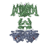 7sizC 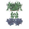 2r9rS S: Starting model for refinement C: citing same article ( |
|---|---|
| Similar structure data | Similarity search - Function & homology  F&H Search F&H Search |
- Links
Links
- Assembly
| Deposited unit | 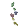
| ||||||||||||||||||
|---|---|---|---|---|---|---|---|---|---|---|---|---|---|---|---|---|---|---|---|
| 1 | 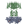
| ||||||||||||||||||
| 2 | 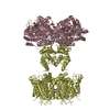
| ||||||||||||||||||
| Unit cell |
| ||||||||||||||||||
| Components on special symmetry positions |
|
- Components
| #1: Protein | Mass: 37353.086 Da / Num. of mol.: 2 Source method: isolated from a genetically manipulated source Source: (gene. exp.)   Komagataella pastoris (fungus) Komagataella pastoris (fungus)References: UniProt: P62483, Oxidoreductases; Acting on the CH-OH group of donors; With NAD+ or NADP+ as acceptor #2: Protein | Mass: 58823.617 Da / Num. of mol.: 2 Mutation: L15H, C31S, C32S, N207Q, W362F, S367T, V377T, C435S, C482S Source method: isolated from a genetically manipulated source Source: (gene. exp.)   Komagataella pastoris (fungus) Komagataella pastoris (fungus)#3: Chemical | #4: Chemical | ChemComp-K / #5: Chemical | ChemComp-O / | Has ligand of interest | Y | |
|---|
-Experimental details
-Experiment
| Experiment | Method:  X-RAY DIFFRACTION / Number of used crystals: 1 X-RAY DIFFRACTION / Number of used crystals: 1 |
|---|
- Sample preparation
| Crystal | Density Matthews: 3 Å3/Da / Density % sol: 59.53 % |
|---|---|
| Crystal grow | Temperature: 293 K / Method: vapor diffusion, hanging drop / pH: 8.5 / Details: 50 mM Tris, pH 8.5, 28-32% PEG400, CHAPS / PH range: 8.3-8.8 |
-Data collection
| Diffraction | Mean temperature: 90 K / Serial crystal experiment: N |
|---|---|
| Diffraction source | Source:  SYNCHROTRON / Site: SYNCHROTRON / Site:  APS APS  / Beamline: 23-ID-D / Wavelength: 1.033 Å / Beamline: 23-ID-D / Wavelength: 1.033 Å |
| Detector | Type: DECTRIS PILATUS 6M / Detector: PIXEL / Date: Mar 5, 2020 |
| Radiation | Monochromator: double crystal / Protocol: SINGLE WAVELENGTH / Monochromatic (M) / Laue (L): M / Scattering type: x-ray |
| Radiation wavelength | Wavelength: 1.033 Å / Relative weight: 1 |
| Reflection | Resolution: 3.32→49.16 Å / Num. obs: 35720 / % possible obs: 99.9 % / Redundancy: 14.9 % / CC1/2: 0.99 / CC star: 0.54 / Net I/σ(I): 7.2 |
| Reflection shell | Resolution: 3.32→3.51 Å / Num. unique obs: 4588 / CC1/2: 0.54 |
-Phasing
| Phasing | Method:  molecular replacement molecular replacement |
|---|
- Processing
| Software |
| ||||||||||||||||||||||||||||||||||||||||||||||||||||||||||||||||||||||||||||||||||||
|---|---|---|---|---|---|---|---|---|---|---|---|---|---|---|---|---|---|---|---|---|---|---|---|---|---|---|---|---|---|---|---|---|---|---|---|---|---|---|---|---|---|---|---|---|---|---|---|---|---|---|---|---|---|---|---|---|---|---|---|---|---|---|---|---|---|---|---|---|---|---|---|---|---|---|---|---|---|---|---|---|---|---|---|---|---|
| Refinement | Method to determine structure:  MOLECULAR REPLACEMENT MOLECULAR REPLACEMENTStarting model: PDB entry 2R9R Resolution: 3.32→49.159 Å / SU ML: 0.56 / Cross valid method: THROUGHOUT / σ(F): 1.33 / Phase error: 31.11 / Stereochemistry target values: ML
| ||||||||||||||||||||||||||||||||||||||||||||||||||||||||||||||||||||||||||||||||||||
| Solvent computation | Shrinkage radii: 0.9 Å / VDW probe radii: 1.11 Å / Solvent model: FLAT BULK SOLVENT MODEL | ||||||||||||||||||||||||||||||||||||||||||||||||||||||||||||||||||||||||||||||||||||
| Displacement parameters | Biso max: 299.21 Å2 / Biso mean: 126.359 Å2 / Biso min: 20 Å2 | ||||||||||||||||||||||||||||||||||||||||||||||||||||||||||||||||||||||||||||||||||||
| Refinement step | Cycle: final / Resolution: 3.32→49.159 Å
| ||||||||||||||||||||||||||||||||||||||||||||||||||||||||||||||||||||||||||||||||||||
| LS refinement shell | Refine-ID: X-RAY DIFFRACTION / Rfactor Rfree error: 0
|
 Movie
Movie Controller


 PDBj
PDBj


