 Movie
Movie Controller

[English] 日本語
 Yorodumi
- PDB-7shj: Crystal structure of Acinetobacter baumannii ZnuA in the metal-fr... -
+ Open data

- Basic information
| Entry | Database: PDB / ID: 7shj | |||||||||
|---|---|---|---|---|---|---|---|---|---|---|
| Title | Crystal structure of Acinetobacter baumannii ZnuA in the metal-free state | |||||||||
 Components Components | Zinc ABC transporter solute-binding protein | |||||||||
 Keywords Keywords | METAL BINDING PROTEIN / zinc / solute-binding protein / ABC transporter / host-pathogen / ZnuA | |||||||||
| Function / homology | : / Periplasmic solute binding protein, ZnuA-like / Zinc-uptake complex component A periplasmic / metal ion transport / metal ion binding / Zinc ABC transporter solute-binding protein Function and homology information Function and homology information | |||||||||
| Biological species |  Acinetobacter baumannii (bacteria) Acinetobacter baumannii (bacteria) | |||||||||
| Method |  X-RAY DIFFRACTION / SYNCHROTRON / MOLECULAR REPLACEMENT / Resolution: 2.13 Å X-RAY DIFFRACTION / SYNCHROTRON / MOLECULAR REPLACEMENT / Resolution: 2.13 Å | |||||||||
 Authors Authors | Luo, Z. / McDevitt, C.A. / Kobe, B. | |||||||||
| Funding support |  Australia, 2items Australia, 2items
| |||||||||
 Citation Citation | Journal: J.Inorg.Biochem. / Year: 2022 Title: Structural and biochemical characterization of Acinetobacter baumannii ZnuA. Authors: Alquethamy, S. / Ganio, K. / Luo, Z. / Hossain, S.I. / Hayes, A.J. / Ve, T. / Davies, M.R. / Deplazes, E. / Kobe, B. / McDevitt, C.A. | |||||||||
| History |
|
- Structure visualization
| Structure viewer | Molecule: MolmilJmol/JSmol |
|---|
- Downloads & links
-Download
| PDBx/mmCIF format | 7shj.cif.gz | 297 KB | Display | PDBx/mmCIF format |
|---|---|---|---|---|
| PDB format | pdb7shj.ent.gz | 234.2 KB | Display | PDB format |
| PDBx/mmJSON format | 7shj.json.gz | Tree view | PDBx/mmJSON format | |
| Others |  Other downloads Other downloads |
-Validation report
| Summary document | 7shj_validation.pdf.gz | 441.2 KB | Display | wwPDB validaton report |
|---|---|---|---|---|
| Full document | 7shj_full_validation.pdf.gz | 446.2 KB | Display | |
| Data in XML | 7shj_validation.xml.gz | 19.6 KB | Display | |
| Data in CIF | 7shj_validation.cif.gz | 27.9 KB | Display | |
| Arichive directory | https://data.pdbj.org/pub/pdb/validation_reports/sh/7shjftp://data.pdbj.org/pub/pdb/validation_reports/sh/7shj | HTTPS FTP |
-Related structure data
| Similar structure data |
|---|
-Links
 PDBj
PDBj- Assembly
| Deposited unit | 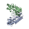
| ||||||||||||
|---|---|---|---|---|---|---|---|---|---|---|---|---|---|
| 1 | 
| ||||||||||||
| 2 | 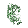
| ||||||||||||
| Unit cell |
|
-Components
| #1: Protein | Mass: 32033.199 Da / Num. of mol.: 2 Source method: isolated from a genetically manipulated source Source: (gene. exp.) Acinetobacter baumannii (bacteria) / Gene: G3N04_06260, G3N12_05260, G3N53_14455, GY141_09205 / Production host: #2: Chemical | ChemComp-NA / |   Mass: 22.990 Da / Num. of mol.: 1 / Source method: obtained synthetically / Formula: Na Mass: 22.990 Da / Num. of mol.: 1 / Source method: obtained synthetically / Formula: Na#3: Water | ChemComp-HOH / |  Mass: 18.015 Da / Num. of mol.: 191 / Source method: isolated from a natural source / Formula: H2O Mass: 18.015 Da / Num. of mol.: 191 / Source method: isolated from a natural source / Formula: H2OHas ligand of interest | N | Has protein modification | Y | |
|---|
-Experimental details
-Experiment
| Experiment | Method: X-RAY DIFFRACTION / Number of used crystals: 1 |
|---|
- Sample preparation
| Crystal | Density Matthews: 1.92 Å3/Da / Density % sol: 35.77 % |
|---|---|
| Crystal grow | Temperature: 293 K / Method: vapor diffusion, hanging drop Details: 0.1 M cirtate pH 7.0, 30% (v/v) polyethylene glycol 6000, and 1 M LiCl |
-Data collection
| Diffraction | Mean temperature: 100 K / Serial crystal experiment: N |
|---|---|
| Diffraction source | Source: SYNCHROTRON / Site: Australian Synchrotron / Beamline: MX2 / Wavelength: 0.954 Å |
| Detector | Type: DECTRIS EIGER X 16M / Detector: PIXEL / Date: Mar 14, 2021 |
| Radiation | Protocol: SINGLE WAVELENGTH / Monochromatic (M) / Laue (L): M / Scattering type: x-ray |
| Radiation wavelength | Wavelength: 0.954 Å / Relative weight: 1 |
| Reflection | Resolution: 2.13→45.55 Å / Num. obs: 27410 / % possible obs: 99.6 % / Redundancy: 2 % / Biso Wilson estimate: 34.29 Å2 / CC1/2: 0.99 / Rmerge(I) obs: 0.05 / Net I/σ(I): 9 |
| Reflection shell | Resolution: 2.13→2.21 Å / Rmerge(I) obs: 0.51 / Num. unique obs: 2701 / CC1/2: 0.57 |
- Processing
| Software |
| |||||||||||||||||||||||||||||||||||||||||||||||||||||||||||||||||||||||||||||
|---|---|---|---|---|---|---|---|---|---|---|---|---|---|---|---|---|---|---|---|---|---|---|---|---|---|---|---|---|---|---|---|---|---|---|---|---|---|---|---|---|---|---|---|---|---|---|---|---|---|---|---|---|---|---|---|---|---|---|---|---|---|---|---|---|---|---|---|---|---|---|---|---|---|---|---|---|---|---|
| Refinement | Method to determine structure: MOLECULAR REPLACEMENT Starting model: model generated by alphaFold2 Resolution: 2.13→45.55 Å / SU ML: 0.2779 / Cross valid method: FREE R-VALUE / σ(F): 1.35 / Phase error: 27.1005 Stereochemistry target values: GeoStd + Monomer Library + CDL v1.2
| |||||||||||||||||||||||||||||||||||||||||||||||||||||||||||||||||||||||||||||
| Solvent computation | Shrinkage radii: 0.9 Å / VDW probe radii: 1.11 Å / Solvent model: FLAT BULK SOLVENT MODEL | |||||||||||||||||||||||||||||||||||||||||||||||||||||||||||||||||||||||||||||
| Displacement parameters | Biso mean: 48.29 Å2 | |||||||||||||||||||||||||||||||||||||||||||||||||||||||||||||||||||||||||||||
| Refinement step | Cycle: LAST / Resolution: 2.13→45.55 Å
| |||||||||||||||||||||||||||||||||||||||||||||||||||||||||||||||||||||||||||||
| Refine LS restraints |
| |||||||||||||||||||||||||||||||||||||||||||||||||||||||||||||||||||||||||||||
| LS refinement shell |
|
