 Movie
Movie Controller


+ Open data

- Basic information
| Entry | Database: PDB / ID: 7sgr | |||||||||
|---|---|---|---|---|---|---|---|---|---|---|
| Title | Structure of hemolysin A secretion system HlyB/D complex | |||||||||
 Components Components |
| |||||||||
 Keywords Keywords | MEMBRANE PROTEIN / hydrolase / transport | |||||||||
| Function / homology |  Function and homology information Function and homology informationtype I protein secretion system complex / protein secretion by the type I secretion system / ABC-type oligopeptide transporter activity / Secretion of toxins / protein secretion / peptidase activity / killing of cells of another organism / ATP hydrolysis activity / proteolysis / ATP binding / plasma membrane Similarity search - Function | |||||||||
| Biological species |  | |||||||||
| Method | ELECTRON MICROSCOPY / single particle reconstruction / cryo EM / Resolution: 2.9 Å | |||||||||
 Authors Authors | Zhao, H. / Chen, J. | |||||||||
| Funding support |  France, France,  United States, 2items United States, 2items
| |||||||||
 Citation Citation | Journal: Cell / Year: 2022 Title: The hemolysin A secretion system is a multi-engine pump containing three ABC transporters. Authors: Hongtu Zhao / James Lee / Jue Chen / Abstract: Type 1 secretion systems (T1SSs) are widespread in pathogenic Gram-negative bacteria, extruding protein substrates following synthesis of the entire polypeptide. The Escherichia coli hemolysin A ...Type 1 secretion systems (T1SSs) are widespread in pathogenic Gram-negative bacteria, extruding protein substrates following synthesis of the entire polypeptide. The Escherichia coli hemolysin A secretion system has long been considered a prototype in structural and mechanistic studies of T1SSs. Three membrane proteins-an inner membrane ABC transporter HlyB, an adaptor protein HlyD, and an outer membrane porin TolC-are required for secretion. However, the stoichiometry and structure of the complex are unknown. Here, cryo-electron microscopy (cryo-EM) structures determined in two conformations reveal that the inner membrane complex is a hetero-dodecameric assembly comprising three HlyB homodimers and six HlyD subunits. Functional studies indicate that oligomerization of HlyB and HlyD is essential for protein secretion and that polypeptides translocate through a canonical ABC transporter pathway in HlyB. Our data suggest that T1SSs entail three ABC transporters, one that functions as a protein channel and two that allosterically power the translocation process. | |||||||||
| History |
|
- Structure visualization
| Structure viewer | Molecule: MolmilJmol/JSmol |
|---|
- Downloads & links
-Download
| PDBx/mmCIF format | 7sgr.cif.gz | 778.1 KB | Display | PDBx/mmCIF format |
|---|---|---|---|---|
| PDB format | pdb7sgr.ent.gz | 633.7 KB | Display | PDB format |
| PDBx/mmJSON format | 7sgr.json.gz | Tree view | PDBx/mmJSON format | |
| Others |  Other downloads Other downloads |
-Validation report
| Arichive directory | https://data.pdbj.org/pub/pdb/validation_reports/sg/7sgrftp://data.pdbj.org/pub/pdb/validation_reports/sg/7sgr | HTTPS FTP |
|---|
-Related structure data
| Related structure data |  25116MC  8dckC M: map data used to model this data C: citing same article ( |
|---|---|
| Similar structure data |

-Links
 PDBj
PDBj

- Assembly
| Deposited unit | 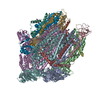
|
|---|---|
| 1 |
|
-Components
| #1: Protein | Mass: 79621.492 Da / Num. of mol.: 6 Source method: isolated from a genetically manipulated source Source: (gene. exp.) #2: Protein | Mass: 40748.895 Da / Num. of mol.: 6 Source method: isolated from a genetically manipulated source Source: (gene. exp.) #3: Chemical | ChemComp-6OU / [( 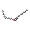  Mass: 717.996 Da / Num. of mol.: 37 / Source method: obtained synthetically / Formula: C39H76NO8P / Comment: phospholipid*YM Mass: 717.996 Da / Num. of mol.: 37 / Source method: obtained synthetically / Formula: C39H76NO8P / Comment: phospholipid*YMHas ligand of interest | N | |
|---|
-Experimental details
-Experiment
| Experiment | Method: ELECTRON MICROSCOPY |
|---|---|
| EM experiment | Aggregation state: PARTICLE / 3D reconstruction method: single particle reconstruction |
- Sample preparation
| Component | Name: Membrane protein complex of HlyB and HlyD / Type: COMPLEX / Entity ID: #1 / Source: RECOMBINANT |
|---|---|
| Molecular weight | Value: 0.81 MDa / Experimental value: NO |
| Source (natural) | Organism: |
| Source (recombinant) | Organism: |
| Buffer solution | pH: 8 |
| Specimen | Embedding applied: NO / Shadowing applied: NO / Staining applied: NO / Vitrification applied: YES |
| Vitrification | Cryogen name: ETHANE |
- Electron microscopy imaging
| Experimental equipment |  Model: Titan Krios / Image courtesy: FEI Company |
|---|---|
| Microscopy | Model: FEI TITAN KRIOS |
| Electron gun | Electron source:  FIELD EMISSION GUN / Accelerating voltage: 300 kV / Illumination mode: FLOOD BEAM FIELD EMISSION GUN / Accelerating voltage: 300 kV / Illumination mode: FLOOD BEAM |
| Electron lens | Mode: BRIGHT FIELD / Nominal defocus max: 3000 nm / Nominal defocus min: 1000 nm |
| Image recording | Electron dose: 70 e/Å2 / Film or detector model: GATAN K2 SUMMIT (4k x 4k) |
- Processing
| CTF correction | Type: PHASE FLIPPING AND AMPLITUDE CORRECTION |
|---|---|
| 3D reconstruction | Resolution: 2.9 Å / Resolution method: FSC 0.143 CUT-OFF / Num. of particles: 136123 / Symmetry type: POINT |
