 Movie
Movie Controller

[English] 日本語
 Yorodumi
- PDB-7sf8: GPR56 (ADGRG1) 7TM domain bound to tethered agonist in complex wi... -
+ Open data

- Basic information
| Entry | Database: PDB / ID: 7sf8 | ||||||
|---|---|---|---|---|---|---|---|
| Title | GPR56 (ADGRG1) 7TM domain bound to tethered agonist in complex with G protein heterotrimer | ||||||
 Components Components |
| ||||||
 Keywords Keywords | MEMBRANE PROTEIN / Adhesion GPCR / GPR56 / ADGRG1 / tethered agonist / stalk / stachel / miniG13 / G13 heterotrimer / G protein / cryoEM | ||||||
| Function / homology |  Function and homology information Function and homology informationcerebral cortex regionalization / cerebral cortex radial glia-guided migration / Rho-activating G protein-coupled receptor signaling pathway / glial limiting end-foot / negative regulation of neuron migration / hematopoietic stem cell homeostasis / regulation of platelet aggregation / layer formation in cerebral cortex / positive regulation of vascular endothelial growth factor signaling pathway / positive regulation of neural precursor cell proliferation ...cerebral cortex regionalization / cerebral cortex radial glia-guided migration / Rho-activating G protein-coupled receptor signaling pathway / glial limiting end-foot / negative regulation of neuron migration / hematopoietic stem cell homeostasis / regulation of platelet aggregation / layer formation in cerebral cortex / positive regulation of vascular endothelial growth factor signaling pathway / positive regulation of neural precursor cell proliferation / extracellular matrix binding / neural precursor cell proliferation / negative regulation of ferroptosis / seminiferous tubule development / positive regulation of Rho protein signal transduction / collagen binding / Rho protein signal transduction / positive regulation of cell adhesion / G protein-coupled receptor activity / brain development / Olfactory Signaling Pathway / Activation of the phototransduction cascade / G beta:gamma signalling through PLC beta / Presynaptic function of Kainate receptors / Thromboxane signalling through TP receptor / G protein-coupled acetylcholine receptor signaling pathway / adenylate cyclase-activating G protein-coupled receptor signaling pathway / Activation of G protein gated Potassium channels / Inhibition of voltage gated Ca2+ channels via Gbeta/gamma subunits / G-protein activation / Prostacyclin signalling through prostacyclin receptor / G beta:gamma signalling through CDC42 / Glucagon signaling in metabolic regulation / G beta:gamma signalling through BTK / Synthesis, secretion, and inactivation of Glucagon-like Peptide-1 (GLP-1) / ADP signalling through P2Y purinoceptor 12 / photoreceptor disc membrane / Sensory perception of sweet, bitter, and umami (glutamate) taste / Glucagon-type ligand receptors / Adrenaline,noradrenaline inhibits insulin secretion / Vasopressin regulates renal water homeostasis via Aquaporins / Glucagon-like Peptide-1 (GLP1) regulates insulin secretion / G alpha (z) signalling events / cellular response to catecholamine stimulus / ADP signalling through P2Y purinoceptor 1 / ADORA2B mediated anti-inflammatory cytokines production / G beta:gamma signalling through PI3Kgamma / adenylate cyclase-activating dopamine receptor signaling pathway / Cooperation of PDCL (PhLP1) and TRiC/CCT in G-protein beta folding / GPER1 signaling / cell migration / Inactivation, recovery and regulation of the phototransduction cascade / cellular response to prostaglandin E stimulus / G-protein beta-subunit binding / heterotrimeric G-protein complex / cell-cell signaling / G alpha (12/13) signalling events / sensory perception of taste / extracellular vesicle / heparin binding / signaling receptor complex adaptor activity / Thrombin signalling through proteinase activated receptors (PARs) / retina development in camera-type eye / GTPase binding / Ca2+ pathway / fibroblast proliferation / High laminar flow shear stress activates signaling by PIEZO1 and PECAM1:CDH5:KDR in endothelial cells / angiogenesis / G alpha (i) signalling events / G alpha (s) signalling events / phospholipase C-activating G protein-coupled receptor signaling pathway / G alpha (q) signalling events / Ras protein signal transduction / Extra-nuclear estrogen signaling / cell surface receptor signaling pathway / cell population proliferation / cell adhesion / G protein-coupled receptor signaling pathway / membrane raft / negative regulation of cell population proliferation / lysosomal membrane / GTPase activity / synapse / protein-containing complex binding / signal transduction / extracellular exosome / membrane / plasma membrane / cytoplasm / cytosol Similarity search - Function | ||||||
| Biological species |  Homo sapiens (human) Homo sapiens (human) | ||||||
| Method | ELECTRON MICROSCOPY / single particle reconstruction / cryo EM / Resolution: 2.7 Å | ||||||
 Authors Authors | Barros-Alvarez, X. / Panova, O. / Skiniotis, G. | ||||||
| Funding support | 1items
| ||||||
 Citation Citation | Journal: Nature / Year: 2022 Title: The tethered peptide activation mechanism of adhesion GPCRs. Authors: Ximena Barros-Álvarez / Robert M Nwokonko / Alexander Vizurraga / Donna Matzov / Feng He / Makaía M Papasergi-Scott / Michael J Robertson / Ouliana Panova / Eliane Hadas Yardeni / Alpay B ...Authors: Ximena Barros-Álvarez / Robert M Nwokonko / Alexander Vizurraga / Donna Matzov / Feng He / Makaía M Papasergi-Scott / Michael J Robertson / Ouliana Panova / Eliane Hadas Yardeni / Alpay B Seven / Frank E Kwarcinski / Hongyu Su / Maria Claudia Peroto / Justin G Meyerowitz / Moran Shalev-Benami / Gregory G Tall / Georgios Skiniotis /   Abstract: Adhesion G-protein-coupled receptors (aGPCRs) are characterized by the presence of auto-proteolysing extracellular regions that are involved in cell-cell and cell-extracellular matrix interactions. ...Adhesion G-protein-coupled receptors (aGPCRs) are characterized by the presence of auto-proteolysing extracellular regions that are involved in cell-cell and cell-extracellular matrix interactions. Self cleavage within the aGPCR auto-proteolysis-inducing (GAIN) domain produces two protomers-N-terminal and C-terminal fragments-that remain non-covalently attached after receptors reach the cell surface. Upon dissociation of the N-terminal fragment, the C-terminus of the GAIN domain acts as a tethered agonist (TA) peptide to activate the seven-transmembrane domain with a mechanism that has been poorly understood. Here we provide cryo-electron microscopy snapshots of two distinct members of the aGPCR family, GPR56 (also known as ADGRG1) and latrophilin 3 (LPHN3 (also known as ADGRL3)). Low-resolution maps of the receptors in their N-terminal fragment-bound state indicate that the GAIN domain projects flexibly towards the extracellular space, keeping the encrypted TA peptide away from the seven-transmembrane domain. High-resolution structures of GPR56 and LPHN3 in their active, G-protein-coupled states, reveal that after dissociation of the extracellular region, the decrypted TA peptides engage the seven-transmembrane domain core with a notable conservation of interactions that also involve extracellular loop 2. TA binding stabilizes breaks in the middle of transmembrane helices 6 and 7 that facilitate aGPCR coupling and activation of heterotrimeric G proteins. Collectively, these results enable us to propose a general model for aGPCR activation. | ||||||
| History |
|
- Structure visualization
| Structure viewer | Molecule: MolmilJmol/JSmol |
|---|
- Downloads & links
-Download
| PDBx/mmCIF format | 7sf8.cif.gz | 160.5 KB | Display | PDBx/mmCIF format |
|---|---|---|---|---|
| PDB format | pdb7sf8.ent.gz | 123.5 KB | Display | PDB format |
| PDBx/mmJSON format | 7sf8.json.gz | Tree view | PDBx/mmJSON format | |
| Others |  Other downloads Other downloads |
-Validation report
| Summary document | 7sf8_validation.pdf.gz | 673.6 KB | Display | wwPDB validaton report |
|---|---|---|---|---|
| Full document | 7sf8_full_validation.pdf.gz | 677.1 KB | Display | |
| Data in XML | 7sf8_validation.xml.gz | 33.5 KB | Display | |
| Data in CIF | 7sf8_validation.cif.gz | 51 KB | Display | |
| Arichive directory | https://data.pdbj.org/pub/pdb/validation_reports/sf/7sf8ftp://data.pdbj.org/pub/pdb/validation_reports/sf/7sf8 | HTTPS FTP |
-Related structure data
| Related structure data |  25077MC 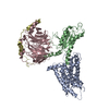 7sf7C M: map data used to model this data C: citing same article ( |
|---|---|
| Similar structure data |
-Links
 PDBj
PDBj


- Assembly
| Deposited unit | 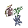
|
|---|---|
| 1 |
|
-Components
| #1: Protein | Mass: 34798.094 Da / Num. of mol.: 1 Fragment: 7TM domain with activation paptide (UNP residues 383-687) Source method: isolated from a genetically manipulated source Source: (gene. exp.) Homo sapiens (human) / Gene: ADGRG1, GPR56, TM7LN4, TM7XN1, UNQ540/PRO1083 / Production host:   Spodoptera frugiperda (fall armyworm) / References: UniProt: Q9Y653 Spodoptera frugiperda (fall armyworm) / References: UniProt: Q9Y653 |
|---|---|
| #2: Protein | Mass: 26574.400 Da / Num. of mol.: 1 Source method: isolated from a genetically manipulated source Source: (gene. exp.) Homo sapiens (human) / Production host: Spodoptera frugiperda (fall armyworm) |
| #3: Protein | Mass: 37416.930 Da / Num. of mol.: 1 Source method: isolated from a genetically manipulated source Source: (gene. exp.) Homo sapiens (human) / Gene: GNB1 / Production host: Spodoptera frugiperda (fall armyworm) / References: UniProt: P62873 |
| #4: Protein | Mass: 7861.143 Da / Num. of mol.: 1 Source method: isolated from a genetically manipulated source Source: (gene. exp.) Homo sapiens (human) / Gene: GNG2 / Production host: Spodoptera frugiperda (fall armyworm) / References: UniProt: P59768 |
| Has protein modification | Y |
-Experimental details
-Experiment
| Experiment | Method: ELECTRON MICROSCOPY |
|---|---|
| EM experiment | Aggregation state: PARTICLE / 3D reconstruction method: single particle reconstruction |
- Sample preparation
| Component | Name: GPR56 (ADGRG1) 7TM domain bound to tethered agonist in complex with mini-G13 protein Type: COMPLEX / Entity ID: all / Source: RECOMBINANT |
|---|---|
| Molecular weight | Experimental value: NO |
| Source (natural) | Organism: Homo sapiens (human) |
| Source (recombinant) | Organism: Spodoptera frugiperda (fall armyworm) |
| Buffer solution | pH: 7.5 |
| Specimen | Conc.: 7.5 mg/ml / Embedding applied: NO / Shadowing applied: NO / Staining applied: NO / Vitrification applied: YES |
| Specimen support | Grid type: UltrAuFoil R1.2/1.3 |
| Vitrification | Instrument: FEI VITROBOT MARK II / Cryogen name: ETHANE |
- Electron microscopy imaging
| Experimental equipment |  Model: Titan Krios / Image courtesy: FEI Company |
|---|---|
| Microscopy | Model: FEI TITAN KRIOS |
| Electron gun | Electron source:  FIELD EMISSION GUN / Accelerating voltage: 300 kV / Illumination mode: FLOOD BEAM FIELD EMISSION GUN / Accelerating voltage: 300 kV / Illumination mode: FLOOD BEAM |
| Electron lens | Mode: BRIGHT FIELD / Calibrated magnification: 55000 X / Cs: 2.7 mm / C2 aperture diameter: 70 µm |
| Specimen holder | Specimen holder model: FEI TITAN KRIOS AUTOGRID HOLDER |
| Image recording | Electron dose: 1.07 e/Å2 / Film or detector model: GATAN K3 BIOQUANTUM (6k x 4k) |
- Processing
| EM software |
| ||||||||||||||||||||||||
|---|---|---|---|---|---|---|---|---|---|---|---|---|---|---|---|---|---|---|---|---|---|---|---|---|---|
| CTF correction | Type: PHASE FLIPPING AND AMPLITUDE CORRECTION | ||||||||||||||||||||||||
| Symmetry | Point symmetry: C1 (asymmetric) | ||||||||||||||||||||||||
| 3D reconstruction | Resolution: 2.7 Å / Resolution method: FSC 0.143 CUT-OFF / Num. of particles: 541279 / Symmetry type: POINT |
