 Movie
Movie Controller

[English] 日本語
 Yorodumi
- PDB-7sdd: Structure of the PTP-like myo-inositol phosphatase from Legionell... -
+ Open data

- Basic information
| Entry | Database: PDB / ID: 7sdd | ||||||
|---|---|---|---|---|---|---|---|
| Title | Structure of the PTP-like myo-inositol phosphatase from Legionella pneumophila str. Paris in complex with myo-inositol-(1,3,4,5)-tetrakisphosphate | ||||||
 Components Components | Myo-inositol phosphohydrolase | ||||||
 Keywords Keywords | HYDROLASE / Phytase / PTP fold / myo-inositol phosphate / IPase | ||||||
| Function / homology |  Function and homology information Function and homology informationprotein tyrosine phosphatase activity, metal-dependent / histone H2AXY142 phosphatase activity / non-membrane spanning protein tyrosine phosphatase activity / protein-tyrosine-phosphatase Similarity search - Function | ||||||
| Biological species |  Legionella pneumophila str. Paris (bacteria) Legionella pneumophila str. Paris (bacteria) | ||||||
| Method |  X-RAY DIFFRACTION / MOLECULAR REPLACEMENT / Resolution: 1.85 Å X-RAY DIFFRACTION / MOLECULAR REPLACEMENT / Resolution: 1.85 Å | ||||||
 Authors Authors | Cleland, C.P. / Mosimann, S.C. | ||||||
| Funding support |  Canada, 1items Canada, 1items
| ||||||
 Citation Citation | Journal: To Be Published Title: Structure of the PTP-like myo-inositol phosphatase from Legionella pneumophila str. Paris in complex with myo-inositol-(1,3,4,5)-tetrakisphosphate Authors: Cleland, C.P. / Mosimann, S.C. | ||||||
| History |
|
- Structure visualization
| Structure viewer | Molecule: MolmilJmol/JSmol |
|---|
- Downloads & links
-Download
| PDBx/mmCIF format | 7sdd.cif.gz | 163.5 KB | Display | PDBx/mmCIF format |
|---|---|---|---|---|
| PDB format | pdb7sdd.ent.gz | 102.8 KB | Display | PDB format |
| PDBx/mmJSON format | 7sdd.json.gz | Tree view | PDBx/mmJSON format | |
| Others |  Other downloads Other downloads |
-Validation report
| Arichive directory | https://data.pdbj.org/pub/pdb/validation_reports/sd/7sddftp://data.pdbj.org/pub/pdb/validation_reports/sd/7sdd | HTTPS FTP |
|---|
-Related structure data
| Related structure data |  4tvvS S: Starting model for refinement |
|---|---|
| Similar structure data |
-Links
 PDBj
PDBj


- Assembly
| Deposited unit | 
| ||||||||||||
|---|---|---|---|---|---|---|---|---|---|---|---|---|---|
| 1 | 
| ||||||||||||
| Unit cell |
|
-Components
| #1: Protein | Mass: 36026.867 Da / Num. of mol.: 1 / Mutation: C231S Source method: isolated from a genetically manipulated source Source: (gene. exp.) Legionella pneumophila str. Paris (bacteria)Gene: hopD2, NCTC12000_03020 / Plasmid: pET28b / Production host: References: UniProt: A0A378K9X8, protein-tyrosine-phosphatase |
|---|---|
| #2: Chemical | ChemComp-4IP / 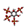  Mass: 500.075 Da / Num. of mol.: 1 / Source method: obtained synthetically / Formula: C6H16O18P4 / Feature type: SUBJECT OF INVESTIGATION Mass: 500.075 Da / Num. of mol.: 1 / Source method: obtained synthetically / Formula: C6H16O18P4 / Feature type: SUBJECT OF INVESTIGATION |
| #3: Water | ChemComp-HOH /  Mass: 18.015 Da / Num. of mol.: 453 / Source method: isolated from a natural source / Formula: H2O Mass: 18.015 Da / Num. of mol.: 453 / Source method: isolated from a natural source / Formula: H2O |
| Has ligand of interest | Y |
| Has protein modification | Y |
-Experimental details
-Experiment
| Experiment | Method: X-RAY DIFFRACTION / Number of used crystals: 1 |
|---|
- Sample preparation
| Crystal | Density Matthews: 2.59 Å3/Da / Density % sol: 52.61 % |
|---|---|
| Crystal grow | Temperature: 293 K / Method: vapor diffusion, sitting drop / pH: 7.4 Details: 100 mM Tris HCl (7.4), 12% PEG 4000, 40 mM magnesium chloride |
-Data collection
| Diffraction | Mean temperature: 100 K / Serial crystal experiment: N |
|---|---|
| Diffraction source | Source: SEALED TUBE / Type: RIGAKU MICROMAX-003 / Wavelength: 1.5418 Å |
| Detector | Type: DECTRIS PILATUS 200K / Detector: PIXEL / Date: Jul 22, 2021 |
| Radiation | Protocol: SINGLE WAVELENGTH / Monochromatic (M) / Laue (L): M / Scattering type: x-ray |
| Radiation wavelength | Wavelength: 1.5418 Å / Relative weight: 1 |
| Reflection | Resolution: 1.85→25.84 Å / Num. obs: 32744 / % possible obs: 99.2 % / Redundancy: 4.6 % / Biso Wilson estimate: 15.2 Å2 / Rmerge(I) obs: 0.093 / Net I/σ(I): 17.23 |
| Reflection shell | Resolution: 1.85→1.92 Å / Rmerge(I) obs: 0.344 / Mean I/σ(I) obs: 2.5 / Num. unique obs: 3278 |
- Processing
| Software |
| |||||||||||||||||||||||||||||||||||||||||||||||||||||||||||||||||||||||||||||||||||||||||||||||||||||||||
|---|---|---|---|---|---|---|---|---|---|---|---|---|---|---|---|---|---|---|---|---|---|---|---|---|---|---|---|---|---|---|---|---|---|---|---|---|---|---|---|---|---|---|---|---|---|---|---|---|---|---|---|---|---|---|---|---|---|---|---|---|---|---|---|---|---|---|---|---|---|---|---|---|---|---|---|---|---|---|---|---|---|---|---|---|---|---|---|---|---|---|---|---|---|---|---|---|---|---|---|---|---|---|---|---|---|---|
| Refinement | Method to determine structure: MOLECULAR REPLACEMENT Starting model: 4TVV Resolution: 1.85→25.84 Å / SU ML: 0.1798 / Cross valid method: FREE R-VALUE / σ(F): 1.33 / Phase error: 20.2618 Stereochemistry target values: GeoStd + Monomer Library + CDL v1.2
| |||||||||||||||||||||||||||||||||||||||||||||||||||||||||||||||||||||||||||||||||||||||||||||||||||||||||
| Solvent computation | Shrinkage radii: 0.9 Å / VDW probe radii: 1.11 Å / Solvent model: FLAT BULK SOLVENT MODEL | |||||||||||||||||||||||||||||||||||||||||||||||||||||||||||||||||||||||||||||||||||||||||||||||||||||||||
| Displacement parameters | Biso mean: 19.62 Å2 | |||||||||||||||||||||||||||||||||||||||||||||||||||||||||||||||||||||||||||||||||||||||||||||||||||||||||
| Refinement step | Cycle: LAST / Resolution: 1.85→25.84 Å
| |||||||||||||||||||||||||||||||||||||||||||||||||||||||||||||||||||||||||||||||||||||||||||||||||||||||||
| Refine LS restraints |
| |||||||||||||||||||||||||||||||||||||||||||||||||||||||||||||||||||||||||||||||||||||||||||||||||||||||||
| LS refinement shell |
|
